Mitochondria is a plural term. The sole form of mitochondria is the mitochondrion. The Greek word ‘Mito’ means thread and ‘Condrion’ means granule. So we can say, mitochondria are thread-like or granular structures. They are found in the eukaryotic cells. We know them as the cell powerhouse or the cell powerplant. It produces ATP (Adenosine triphosphate) by the oxidation process. The name of the process is oxidative phosphorylation. Actually, it is the main role of a mitochondrion. This is kind of an exergonic reaction. Exergonic reaction means the reactions that release energy.
Now, there may arise a question at this point. Certainly, mitochondria produce energy for the eukaryotic cells. But what about the prokaryotic ones? Well, the enzymes of the plasma membrane, carry out the prokaryotic cell’s oxidation of organic materials.
Kolliker first observed mitochondria in the muscle cells of insects in 1880. He named them Sarcosomes. After that, Carl Benda coined the name Mitochondria between 1897-98.
Best safe and secure cloud storage with password protection
Get Envato Elements, Prime Video, Hotstar and Netflix For Free
Best Money Earning Website 100$ Day
#1 Top ranking article submission website
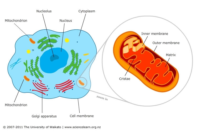
Size & Shape
The size and shape of mitochondria vary from cell to cell. Such as rod-shaped, fibrous, and round. The helical shape is found in sperm cells. Star-shaped mitochondria are found in the pancreatic cells and muscle tissue.
A mitochondrion is about 0.5 to 1.0 µm in diameter and 2 to 8 µm in length. But it varies from cell to cell. It is the smallest in Yeast cells. However, in exocrine cells of the mammalian pancreas, they are about 10 μm long. And in the oocytes of an amphibian called Rana pipiens mitochondria are 20 to 40 µm long.
Number of Mitochondria
The number of mitochondria varies from cell to cell and from species to species. For example, a few algae and some protozoans have only one mitochondrion. Because their number is related to the activity, age, and type of the cell. So, growing, dividing, and actively synthesizing cells contain more mitochondria than the other cells. For instance in Amoeba (Chaos chaos), there may be as many as 50,000 mitochondria. In the case of rat liver cells, these are few in number. About 1000 to 1600. There are as many as 3, 00,000 mitochondria in some oocytes. But matured RBC and prokaryotic cells don’t have any mitochondria at all.
Distribution
Mitochondria lie freely in the cytoplasm. Basically, they own the power of free movement. Sometimes, they may look like threads. In some cells, they can move freely. Moreover, they can carry ATP where needed. But, they are settled permanently near the region where the cell needs more energy. Rod and cone cells of the retina have mitochondria placed in the inner segment. Whereas the cells of kidney tubules have mitochondria in the folds of basal regions near the plasma membrane. And in neurons, they are located in the transmitting region of impulse. During cell division, they get centered around the spindle.
Physical Structure
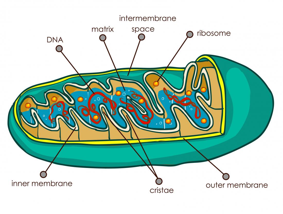
There are 6 parts of mitochondria. We can see them under an electron microscope. They are-
- Outer membrane
- Inner membrane
- Intermembrane space
- Cristae
- Oxysome &
- Matrix
Outer membrane
This membrane is alike the plasma membrane in molecular structure. It is 60-70Å thick. It has two layers of phospholipid molecules. Phospholipid layers are sandwiched between two layers of protein molecules. This outer membrane is smooth and permeable to most small molecules. It consists of about 50% lipid. And includes a large amount of cholesterol. There are different kinds of enzymes. But the mass of protein is really poor.
Inner membrane
The inner membrane is also like the plasma membrane. It is 60-70Å thick. And it has two layers of phospholipid molecules. Those are also sandwiched between two layers of protein molecules. The inner membrane is selectively permeable. So, it controls the flow of materials into and out of the mitochondrion. The inner layer is also rich in enzyme and carrier proteins permease. It has a very high protein/lipid ratio (about 4:1 by weight). But it lacks cholesterol.
Intermembrane space
The mitochondrial intermembrane space is the space within the outer membrane and the inner membrane. The width of the space is about 80Å. Some other names are the outer chamber and, peri mitochondrial space. It looks similar to the cytosol because the space contains clear homogeneous fluids.
Cristae
The inner mitochondrial membrane has many folds in it. These finger-like folds are called Cristae. Cristae expand the surface area of the inner mitochondrial membrane. In addition, it enhances its ability to produce ATP. The pattern of cristae has different characteristic styles in different cells. And they also vary in number. Though the active cells may have numerous cristae. But the idle cells may have only a few.
Oxysomes
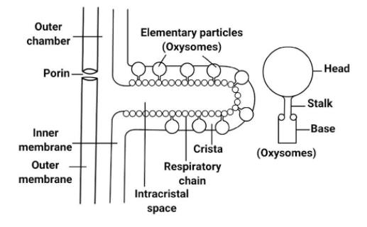
Cristae have many small round figures attached with them. So these are the inner membrane subunits. These are also called elementary particles (EP) or oxysomes. Oxysome has three parts. They are-
- A rounded headpiece or F1 sub-unit
- A short stalk and,
- A base piece or F0 sub-unit.
There may be 100,000 to 1000,000 oxysomes in a mitochondrion.
Matrix
The space inside the inner membrane and between the cristae is called the matrix. It is filled with a gel-like element. This is also called the mitochondrial matrix. It has proteins, lipids, ribosomes, RNA, and one or two DNA molecules. Moreover, it has certain fibrils, crystals, and dense granules. The matrix contains a high density of a mixture. Because the mix consists of hundreds of enzymes. So the matrix is essential for ATP synthesis.
Chemical Composition
The mitochondrion carries ¾ protein and ¼ lipid. It has-
¾ Protein |
¼ Lipid |
|
70 types enzymes 14 types co-enzyme NAD Flavoprotein and Cytochrome |
Cholesterol (25%) Phospholipid (70%) Triglycerides and Fatty acids (5%) |
Functions of Mitochondria
Powerhouse: Firstly, cell respiration takes place in mitochondria. And it produces a lot of energy (ATP). So mitochondria are called the powerhouse of the cell.
Provider for synthesis: Secondly, it provides intermediates for the synthesis of major biomolecules. Such as chlorophyll, cytochromes, steroids, etc.
Amino acids: Thirdly it takes part in the formation of some amino acids.
Storage: Further, mitochondria actively stock calcium ions. It also controls the density of calcium ions in the cytoplasm. In the same vain storing and releasing them is another important work of it.
Enzymes: Mitochondria also hold enzymes and co-enzymes for respiration.
Respiration: Moreover, the Krebs cycle occurs in the matrix. And the electron transport system occurs in the membrane of Mitochondria. So the importance is mitochondria for cell respiration is tremendous.
Apoptosis: In addition, mitochondria function in stress response. It is also responsible for apoptosis. Apoptosis means the process of programmed cell death.
Others: It also plays a key role in photorespiration and cytoplasmic male sterility.
Don’t forget to check out: Biogenesis of Mitochondria.
References
- Lecture of Chandan Kumar Dash Sir (Lecturer, Department of Botany, University of Dhaka)
 Plantlet The Blogging Platform of Department of Botany, University of Dhaka

I’m often to blogging and i really appreciate your content. The article has actually peaks my interest. I’m going to bookmark your web site and maintain checking for brand spanking new information.
I am delighted to know that our blogs are making your interest grow better. Do visit whenever you want to learn more. Have a great day.
very informative articles or reviews at this time.
Thank you so much for your appreciation. Means a lot.
Your article gave me a lot of inspiration, I hope you can explain your point of view in more detail, because I have some doubts, thank you.
Thank you for your appreciation. Please let me know which doubts you are facing in details.