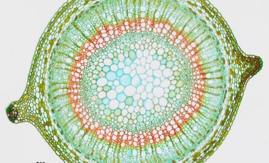
The part of the axis of the plant which is usually ascending and aerial in nature, and also bears the leaves and reproductive structures is called the stem. The stem together with the leaves which it bears constitute the shoot. It is composed of three tissue systems that include the epidermis, vascular, and ground tissues, all of which are made from simple cell types. In this article, the Primary Structure of Stem in Monocot and Dicot plants will be discussed explicitly.
Primary Structure of Dicot Stem
In young dicotyledonous stems, there are three distinct regions – the epidermis, the cortex, and the stele.
These are described below:
Epidermis
The epidermis consists of a single layer of cells and is the outermost layer of the stem.
- It contains stomata and produces various types of trichomes.
- The outer walls are greatly thickened and heavy cutinized.
- The cells are compactly arranged and do not possess intercellular spaces.
- In transverse section, the cells appear almost rectangular.
Function
- It serves mainly for restricting the rate of transpiration
- It protects the underlying tissue from mechanical injury and from disease-producing organisms.
Cortex
The region that lies next to the epidermis is the cortex.
- The innermost layer of the cortex is the endodermis, known also as the starch-sheath.
- It consists of a single layer of cells that surrounds the stele and contains numerous starch grains.
- Frequently it is most easily distinguishable from the surrounding tissue by the presence of these starch grains.
Best safe and secure cloud storage with password protection
Get Envato Elements, Prime Video, Hotstar and Netflix For Free
Best Money Earning Website 100$ Day
#1 Top ranking article submission website
The part of the cortex situated between the epidermis and endodermis is generally divided into two regions:
- An Outer zone of collenchyma cells.
- An Inner zone of parenchyma cells.
Collenchyma
On the inside of the epidermis, there is usually band of collenchyma.
- The cells of the collenchyma are modified parenchyma cells with cellulose walls thickened at the angles where three or more cells are in resembles parenchyma in being alive and in having a moderate amount of protoplasm.
- Collenchyma cells of stem sometimes contain chloroplast.
Function
- The chief function of collenchyma cells is to serve as strengthening material in succulent organs which do not develop much woody tissue, or in the soft young parts of woody plants before stronger tissues have been developed.
- They are specially fitted for giving strength to young, growing organs, since the thickened parts of the walls have considerable rigidity, while the thinner parts allow for an exchange of materials between the cells and for the cells.
- Collenchyma cells carry on photosynthesis when they contain chloroplasts.
Parenchyma
Parenchyma cells are generally regular in shape, have comparatively thin walls, and are not greatly elongated in any direction.
- They are living cells and contain a moderate amount of protoplasm.
- When they are exposed to light they develop chloroplasts and are known as chlorenchyma cells.
- Chlorenchyma cells are thus only a special kind of parenchyma cells.
- The parenchyma cells in the cortex of a stem are near enough to the light so that some or all of them develop chloroplasts and perform photosynthesis.
Function
- The function of parenchyma cells is important in succulent stems and in the young parts of the stems and woody plants before strong mechanical tissues have been developed.
- The parenchyma cells serve for the slow conduction of water and food.
- In the case of cortex of stem, it becomes evident that the water which is received by the collenchyma and the epidermis must be conducted through the parenchyma.
- The turgid parenchyma cells frequently help in rigidity to an organ.
- The parenchyma is the special storage tissue of plants.
Sclerenchyma
- Sclerenchyma cells are found in the cortex of some stems.
There are two varieties of these sclerenchyma cells
- Short or irregularly shaped cells, known as stone cells
- Sclerenchyma Fibres
- Sclerenchyma fibres are long, thick-walled dead cells
- Sclerids have been reported from the cortex of many water plants (e.g., Limnanthemum, Nymphaea, etc.).
Function
- Sclerenchyma fibres serve as strengthening material.
- Stone cells give stiffness to the cortex.
Endodermis
The innermost layer of the cortex is the endodermis consisting of barrel-shaped, elongated, compact cells, having no intercellular space among them.
- Usually, the cells contain starch grains and thus the endodermis may be termed as starch sheath.
- It surrounds stele.
Stele
The part of the stem inside of the cortex is known as the stele. The stele consists of three general regions:
- Pericycle
- Vascular bundle region
- Pith
These three regions are described below:
1. Pericycle
The region between the vascular bundles and the cortex is known as the pericycle.
- It is generally composed of parenchyma and sclerenchyma cells but sclerenchyma cells may be absent.
- The sclerenchyma may occur as separate patches or as a continuous ring in the outer part of the pericycle, forming a sharp line of demarcation between the stele and the cortex.
- The sclerenchyma cells in the pericyle are like other sclerenchyma cells in being long, thick-walled dead cells which serve as strengthing material.
2. Vascular Bundle
The vascular bundles as seen in cross-section, are arranged in the general form of a broken ring. Each vascular bundle consists of three parts.
- Xylem
• The nearest centre of the stem contained thick-walled cells and is known as xylem. - Phloem
• The peripheral portion of the bundle is composed of thin-walled cells called phloem. - Cambium
• The xylem and phloem are separated by a cambium layer, which is composed of meristematic cells.
The xylem which is formed before the activity of the cambium has begun to produce xylem and phloem cells is called primary xylem. It is composed of two parts:
- Protoxylem
1. The xylem formed first is nearest the center of the stem is called protoxylem.
- Metaxylem
2. The more peripheral part of primary xylem is known as metaxylem.
3. Pith
In a dicotyledonous plant, the centre of the stem is composed of thin-walled parenchyma cells and is known as the pith.
- The cells have distinct intercellular spaces.
Pith Ray
The vascular bundles are separated from each other by radial rows of parenchyma cells known as pith rays.
- The pith ray cells are usually elongated in a radial direction.
Function
- They serve primarily for the conduction of food and water radially in the stem and for the storage of food.

Primary Structure of Monocot Stem
The monocotyledonous stems are similar to dicotyledonous stems in having an epidermis, a cortex and a stele.
- The cortex may be well developed and sharply marked off from the stele or it may be quite narrow and inconspicuous.
- It is the structure and arrangement of bundles that monocotyledonous stems, differ markedly from dicotyledonous stems.
Stele
- Vascular bundles of monocotyledonous stems are usually scattered throughout the stele including the pith so that there is no distinction between pith and pith rays.
- Sometimes the center of the stele is free from vascular bundles and is occupied by parenchyma cells which dry up and disappear at an early stage resulting in a hollow stem, as in most grasses.
Vascular Bundle
- The vascular bundles of monocotyledonous stems are like those of dicotyledonous stems in consisting of xylem towards the centre of the stele and phloem towards the periphery.
- The vascular bundles of monocotyledonous stem do not possess a cambium layer which is found in dicotyledonous stems. This means that monocotyledonous stems usually do not have secondary thickening.
- Each bundle remains more or less completely surrounded by a sheath of sclerenchyma cells, the bundle sheath which is particularly well developed on the sides towards the centre and towards the periphery of the stem.
- The phloem is made up mostly of sieve tubes, companion cells, and xylem of vessel and woody parenchyma.

Characteristics of Monocot Stem
The most distinctive and characteristic features of the monocot stem are as follows:
- The Vascular bundles are many.
- The stele is broken up into bundles. The vascular bundles are lying scattered in the ground tissue of the axis.
- The endodermis is not found. The cortex, pericycle, and pith are not differentiated because of the presence of scattered bundles throughout the axis.
- The vascular bundles are collateral and closed. The secondary growth of usual type is lacking but vestiges of cambial activity in bundles may present in the plant body.
- Leaf trace bundles are numerous. The leaf traces when enter the stem, penetrate deeply. The median traces penetrate more deeply than lateral. The bundles are common. Each common bundle somehow or other fuses with other bundles in the due course of time. The anastamoses occur at the nodes.
- Each vascular bundle remains surrounded by a well-developed sclerenchyma sheath.
- The vascular bundle are commonly oval-shaped.
- The phloem is represented by sieve tubes and companion cells only. The phloem parenchyma is not found.
- The pith is not marked out.
- Usually, sclerenchymatous hypodermis is present.
- Usually, epidermal hairs are not present.
References
- Plant Anatomy by B.P. Pandey
Revised By
- Shajneen Jahan Shoily on 28th August 2021.
 Plantlet The Blogging Platform of Department of Botany, University of Dhaka


