The simplest members of the order Marchiantales are found in the family Ricciaceae. There are roughly 140 species in the family. They fall under the Tesselina, Ricciocarpus, and Riccia genera. There are only one species each for the first two. The genus Riccia encompasses the remainder. The Italian botanist F. F. Ricci is honored by the name Riccia.
Classification
Division: Bryophyta
Class: Hepaticopsida
Order: Marchiantales
Family: Ricciaceae
Genus: Riccia
Distribution and Habitat
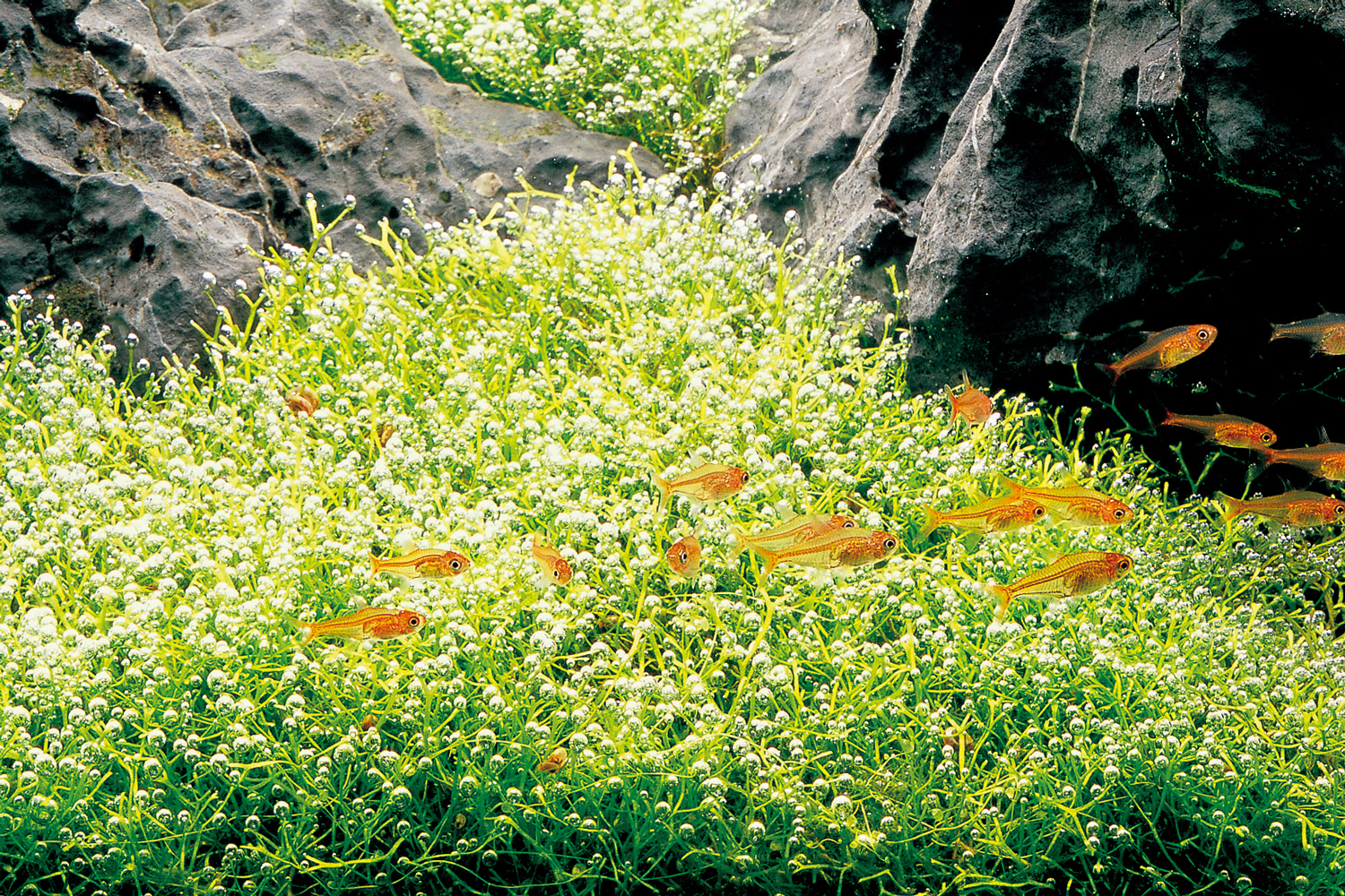
Riccia is a typical liverwort in most cases. The family Ricciaceae is the genus with the broadest distribution. Among all bryophytes, the Riccia species is widespread and can be found anywhere. Numerous species can be found in tropical and subtropical areas. They are visible or growing on the ground or a layer of a damp substrate during the rainy season. There are 138 species total, 137 of which are terrestrial, and just one aquatic species, Riccia fluitans.

The external structure of the plant body
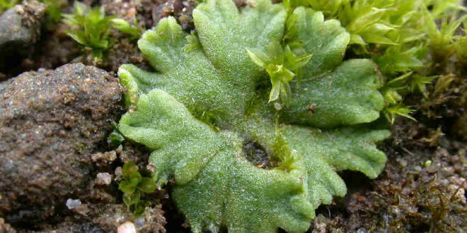
Gametophytic thallus is the main plant body of the Riccia. They typically have dichotomous dorsiventral branches. These branches are very near to one another. Aquatic species are light green, whereas all others are deep green. Dichotomously branching thallus assemble to form the rose-like structure- Rosette. Notches, scales, and rhizoids are important thallus parts.
Notches:
- Apical notch:
- the leading edge of the thallus.
- a slender part.
- thallus development takes place in this section.
- Groove:
- There is a prominent median longitudinal groove on each thallus branch.
- Every branch of the thallus has a groove on the dorsal surface.
- Midrib:
- Green in color, the upper section has a thick midrib.
Scales:
- emerges from the ventral surface.
- situated on the thallus’s edge.
- multicellular one-celled.
- Violet in complexion
- Scales are, close to each other towards- Apex and quite far from each other in Notch
- Scales are present in two rows in the region distant from the apex and one row toward- the apex.
Rhizoids:
- Located at the center of the ventral surface.
- Absorbs water and nutrients from the substrate.
- Two varieties: Tuberculate (with infolding peepings that resemble pegs) and Smooth-walled (no such foldings)
Internal structure (Anatomy)
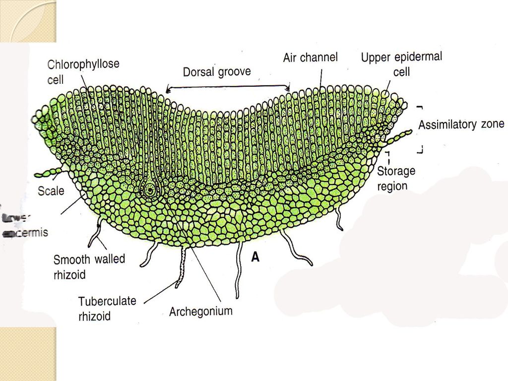
Two separate interior zones are visible in the thallus’ transverse portion under a microscope.
Dorsal zone: A layer with chloroplast which is a photosynthetic zone.
Ventral zone: A colorless parenchyma layer which is a storage region.
Dorsal zone:
Assimilatory filament:
- A cell layer with vertically arranged rows of chloroplasts is present on the dorsal surface (parenchymatous cell rich in the chloroplast).
- It is a photosynthetic area. Carbohydrates are prepared here.
- Loose, green tissue makes up this portion.
Air chamber:
- A midrib area between each cell layer, referred to as an air chamber, is filled with air.
Upper epidermis:
- This is the uppermost single layer of the dorsal surface.
- The epidermis of the thallus is not continuous, however continuous in aquatic species.
- The cells of the upper epidermis are swollen, pear-shaped, and colorless.
Air pores:
- Originates because of discontinuous air pores.
- These air pores aren’t well-defined.
- 4-8 cells build up one pore.
- Works for gaseous exchange.
Ventral zone:
Parenchymatous cell:
- Simple, colorless, and compactly arranged, without any intercellular space.
Epidermis:
- In this region, the single-layered epidermis gives rise to rhizoids (unicellular) and scales (multicellular). Unicellular rhizoids can be of 2 types: Tuberculate and Smooth.
Stored products:
- Filled with starch grains.
- Serve for food and water storage.
Reproduction
Riccia vegetatively reproduces via Fragmentation, Formation of adventitious branches, Persistent growing apices, and formation of tubers.
Sexual reproduction is an oogamous type. Here motile flagellate male gamete is born inside antheridia and the resting non flagellate female gamete is born inside archegonia. Riccia may be of monoecious (e.g. R. gluca) or dioecious ( e.g. R.discolor) type. Sex organs arise in acropetal succession.
Structure of mature Antheridium:
The mature antheridium remains embedded in the antheridial chamber. The antheridium chamber opens by an ostiole on the dorsal side of the thallus. The mature antheridium consists of a few-celled stalk. It also consists of an antheridial proper. It can be rounded or pointed at its apical end. Antheridium is encircled and protected by a sterile single-layered jacket. The mature antheridium contains androcytes within the jacket layer. Each androcytes metamorphose into an antherozoid.

Development of Antheridium:
- The development of Antheridium starts from a superficial antheridial cell, situated on the dorsal surface of the thallus.
- The antheridial initial enlarges in size, becomes papillate, and divides first by transverse division to form an upper outer cell and a lower basal cell. The basal cell remains embedded in the tissue of the thallus, undergoes only a little further development, and forms the embedded portion of the antheridial stalk. The outer cell divides by transverse division to form a filament of 4 cells. The Upper 2 cells of the 4-celled filament are known as primary antheridial cells and the lower 2 cells are known as primary stalk cells.
- Primary stalk cells form the stalk of the antheridium. Primary antheridial cells divide by two successive vertical divisions at right angles to each other to form two tiers of 4 cells each.
- A periclinal division is laid down in both the tiers of four cells and there’s the formation of 8 outer sterile jacket initials and 8 inner androgonial cells.
- Jacket initials divide by several anticlinal divisions to form a single layer of the antheridial jacket. Primary androgonial cells divide by several repeated transverse and vertical divisions resulting in the formation of a large number of small cubical androgonial cells. The last generation of androgonial cells is known as an androcyte mother cell.

Spermatogenesis:
The process of metamorphosis of an androcyte mother cell into an antherozoid is called Spermatogenesis. It completes within a few minutes.
- Each androcyte mother cell divide by a diagonal mitotic division to form two triangular cells called Androcytes. Both the androcytes remain enclosed in the wall of the androcyte mother cell with one separate wall.
- Each androcyte has a prominent nucleus and a small extra nuclear granule called Blepharoplast. It lies near the periphery of the protoplast. The androcyte soon loses its triangular shape and becomes somewhat round or oval. Its blepharoplast elongates into a cord and occupies about two-thirds of its part.
- Simultaneously the nucleus also becomes crescent-shaped, and homogeneous and ultimately comes in contact with the blepharoplast.
- Two large flagella develop from the conspicuously thickened end of the blepharoplast.
- A small part of the cytoplasm, which is not utilized in the formation of flagella may remain attached to the posterior end of the antherozoid as a small vesicle. Each androcyte thus metamorphoses into an antherozoid.
Structure of a mature Archegonium:
Archegonium is a flask-shaped body. It is attached to the thallus by a short stalk. It consisted of an elongated stalk and bulbous venter. The neck is 6-9 cells in height. It consists of 6 vertical columns of cells. They enclose a neck canal. There are 4 cover cells at the top of the neck canal. 4 neck canal cells are found within a neck canal before maturity. They disintegrate into mucilaginous mass on the maturation of the archegonium. Venter is the lower bulbous structure of the Archegonium. This has a single-layered wall around it. It is of 12-20 cells in the perimeter. The venter encloses a venter canal cell and a large egg. The venter canal cell disintegrates on the maturity of the archegonium. Thus only the large egg remains.
Development of Archegonium:
- AArchegonium develops from a superficial cell called archegonial initial, which lies 2 or 3 cells away from the apex of the thallus.
- The archegonial initial divides by a transverse division into 2 cells: Basal c
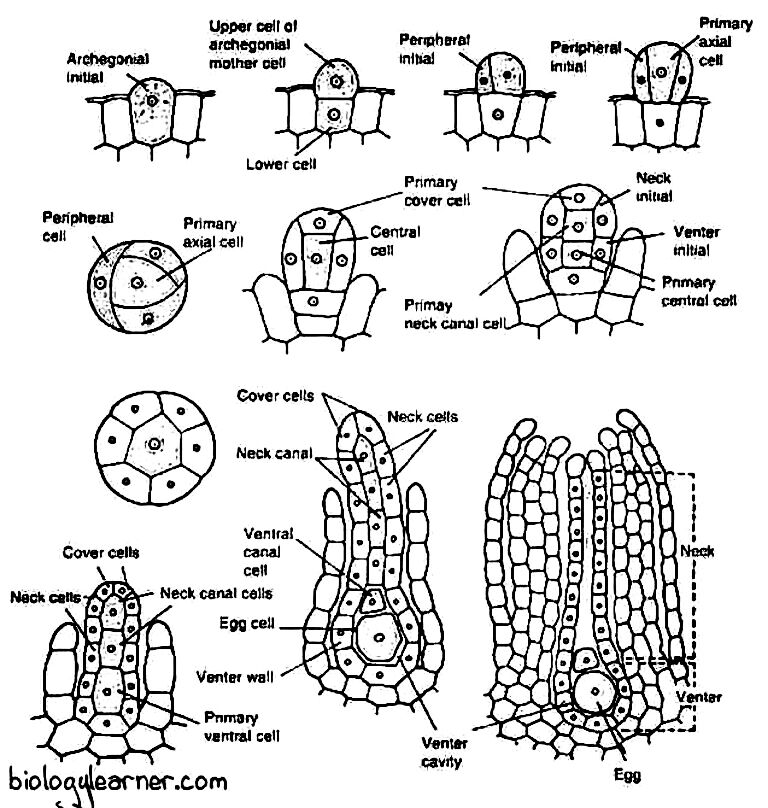
Development of Archegonia. Source: biologylearner.com ell and Outer cell. The basal cell is the lower cell, it produces the lowermost portion of the archegonium. The outer cell is the upper cell, it produces the main body of the archegonium.
- The outer cell divides vertically 3 times, giving rise to 3 peripheral initials. It becomes the primary axial cell. The 3 peripheral cells are situated upon a primary axial cell.
- The 3 peripheral initial divides vertically and form 6 jacket initials. All 6 cells surround the central primary axial cell. Soon after each jacket initial divides by a transverse division producing 6 neck initials and 6 venter initials. They are arranged in 2 tiers. The tier of 6 neck cells is situated above the tier of 6 venter initials.
- Several transverse divisions occur in the neck initials. It gives rise to a neck of archegonium 6-9 cells in length and 6 cells in parameter. The venter initials divide to form the jacket of the venter. The jacket of the venter is 2-20 cells in parameter.
- The primary axial cell divides by an unequal transverse division and produces a small primary cover cell and a central cell. The primary cover cell divides twice vertically. It produces 4 cover cells at the apex of the jacket layer. The central cell divides transversely and produces the primary neck canal cell and primary venter cell.
- The primary neck canal cell divides twice. It gives rise to 4 neck canal cells. They are situated within the neck of the archegonium. The primary venter cell divides by an unequal transverse division and gives rise to a venter canal cell and a large egg. The Venter canal cell is situated over the egg.
The neck canal cells, venter canal cells, and cover cells are separated from each other in mature archegonium.
Fertilization and Syngamy:
- Water is essential for fertilization. Water permits the movement of the antherozoid and helps them to reach the mouth of the neck of the archegonium.
- Neck canal cell in mature archegonia degenerates to form mass mucilage.
- As mass mucilage absorbs water, mucilaginous substances along with proteins and inorganic salts ooze out through the mouth of the archegonium.
- The antherozoids are attracted chemotactically by this substance.
- Antherozoids enter the mouth of the archegonium.
- They travel through the egg and reach near the egg.
- One of the antherozoids penetrates the egg cell and fertilization takes place.

Development of Embryo:
- Formation of calyptra: The zygote secrets a wall around it after fertilization. It nearly fills the cavity of the venter. The venter cells divide periclinally and anticlinally and it becomes two-celled in thickness. Thus form 2 layered calyptra. The developing embryo is situated inside the calyptra.
- Formation of octant: The first division of the zygote is transverse. Thus two equal cells are formed. These 2 cells are called epibasal and hypobasal cells. They divide vertically and 4 celled embryo of quadrant type is formed; it divides by a vertical wall, thus 8 celled embryo is formed, called the octant stage.
- Development of amphithecium and endothecium: Several irregular divisions occur in this stage and give rise to a 20 to 30-celled embryo. Then periclinal division occurs; Thus the embryo is now differentiated into two regions amphithecium and endothecium. Amphithecium is the outer layer, it is protective, and forms the jacket of the capsule. Endotheciu is the inner mass, that divides repeatedly, and gives rise to a mass of sporogenous cells.
- The sporogenous tissue differentiates between sporocyte and nurse cells.
- The nurse cell and their inner jacket cell disintegrate. They give rise to mucilaginous mass. The sporocyte remains suspended in mucilaginous mass, receives nourishment from this, goes through miosis division, and forms spore tetrad. Finally, the walls of the spore mother cells disintegrate. Now they lie free in sporogonium.

Sporophyte characteristics:
- Foot and seta are absent.
- The capsule is globose.
- The capsule wall is one-layered.
- Amphithecium forms the jacket of the capsule.
- Endothecium forms the archesporium.
- Archesporium gives rise to the nurse cells and spore mother cells.
- No special mechanism of dehiscence. Spores are set free after the death of the thallus.
Germination of spore:
Spore is the beginning of gametophytic generation. It has the following processes

of generation:
- The outer layer of the spore, exosporium, and exosporium rupture, and the endosporium protrudes out through the opening and forms a germinal tube.
- Much of the cytoplasm is shifted in the terminal end; transverse walls are laid down at the apex of the tube and a rhizoid comes out near the germinal tube of the spore.
- The distal end of the germinal tube again divides by a parallel wall and 2 cells are formed at the distal end of the germinal tube.
- Both the cells divide vertically twice and give rise to 2 tiers of 4 cells each.
- The 4 cells divide transversely, giving rise to the cylindrical elongated posterior portion of the young gametophyte.
- One cell of the four-celled distal tier acts as an apical cell. It possesses two cutting faces and gives rise to a multicellular young thallus.
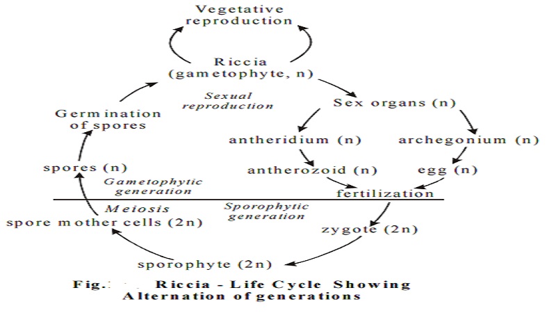
Life-cycle
References
- Botany for degree students: Bryophyta by B.R Vashita, A.K Sinha, and Adarsh Kumar
- Introduction to Bryophytes by Vanderpoorten and Goffinet
 Plantlet The Blogging Platform of Department of Botany, University of Dhaka


I was recommended this website by my cousin. I am not sure whether this post is written by him as nobody else know such detailed about my difficulty. You are wonderful! Thanks!