Megasporangium is the structure of a plant body that contains a female reproductive organ. It can be called an ovule. It consists of nucellus and integument. The ovule can be formed in different sizes and shapes. Many times it changes its form during the course of its development.
Mature megasporangium can be classified under 5 types.
- Orthotropous or atropous
- Anatropous
- Camphylotropous
- Amphitropous
- Hemianatropous
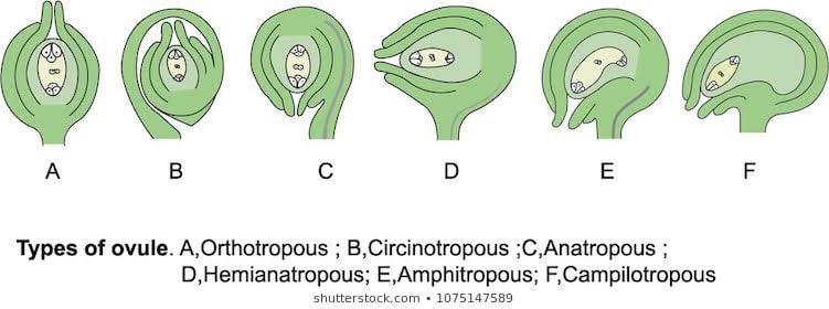
Orthotropous – The micropyle lies directly in line with the hilum. Examples -Polygonaceae, Piperaceae
Anatropous– The ovule becomes completely Inverted so that the micropyle and hilum lie close to each other. For example – all members of Sympetale and several families in monocot and dicot.
Camphylotropous- The ovules are curved. Example – Leguminosae.
Amphitropous- When the curvature of the ovule is more pronounced, it affects the embryo sac, it bents like a horseshoe. Examples- are Alismaceae and Butomaceae.
Hemianatropous- When the integument and nucellus lie at a right angle, for example- Ranunculus, Tulbaghia.
A very peculiar type of ovule is seen in some members of Plumbaginaceae, here the curvature does not stop but rather continues until the ovule turns completely. This type is called Circinotropous. Example- Opuntia.
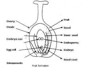
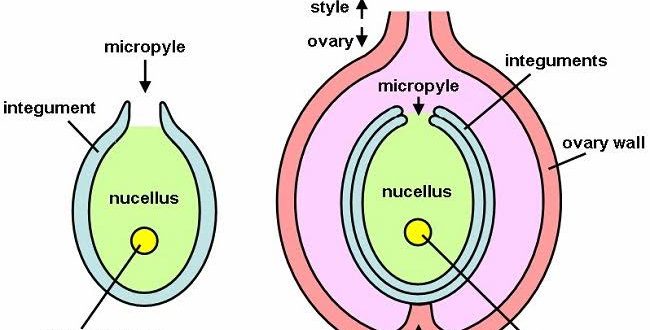
Integument – Nucellus is surrounded by a cover, except in the micropilar region is called the integument. Generally, the ovule has one or two integuments. Most genera in monocot have two integuments.
There is a evidence that in some genera by a fusion of two separate primordia, a single integument has originated.
- In Opuntia a peculiar condition occur where the extremely long funicles surrounds the ovule n looks like a third integument . The 3rd integument can arise by splitting of outer integument , example – Ulmus . But most other cases it is a new structure arising from the base of the ovule , example – Asphodelus . In Euphorbiaceae, 3rd integument arise by a proliferation of the integumentary cells at the micropylar region , called Caruncle.
- In overy if there is one integument, it is called Unitegmic and if there are two integument , it is called Bitegmic. In Sympetale ( 1 petal is present ) flower one integument is present except – Plumbaginales , Primulales. If there is no integument, it is called Ategmic , example – Lauranthus , Santalus .
- In many plants it can be in different structure at young stages . In mature stage it can be absent or remain in a complicated structure .
- Some integument can possess chlorophyll , example – Morigna olifera , Gladiolus communis .
- Stomata can also found in integument , example – Gossypium, Curvularia .
Function
- Normally forms seed coat in mature stage
- Sometimes, supply nutrition
Sometimes remain as a thin layer
Micropyle- It may be formed either by the inner integument, in Centrospermales or by both inner and outer integument , in Pontederiaceae where there is two integuments are present.
When both integument take part in formation , a passage is formed by the outer integument, as a result the micropylar canal has a zigzag outline, example- Melastomaceae . In Ficus, the integumentary cells come in contact with each other so that the canal of micropyle is extremely narrow and imperceptible.
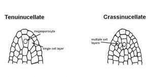
Nucellus- Depending on the development of the nucellus , the ovules can be two type-
Crassinucellate – A well developed parietal tissue is present and the microspore mother cell is separated from the nucellar epidermis by one or several layer . The nucellus may enlarge by an increasing number of parietal cell or by periclinal division, example- Zizyphus . Several members in Salicaceae, Polygonaceae has a beak- shaped nucellus reaching out the micropyle.
Tenuinucellate- The parietal cell is absent and megaspore mother cell lies directly below the necellar epidermis, example- Rubiaceae, Orchidaceae.
When the embroyo sac matures, the cell of nucellus becomes used up.
There is another type of ovule can be found –
Pseudo – crassinucellate – No partial cell is present . The epidermis is divided and form many layer of cell. Example – Nigella damascena
Integument tapetum- Where the nucellus is disorganized, the embryo sac comes in contact with the inner layer of seed coat, they show a pronounced radial elongation and sometimes binucleate . Having the similarities with the anther tapetum, this layer is known as integument tapetum or endothelium . It is a nutritive layer whose main function is to serve as an medium of transportation of food materials from integument to embryo sac . In later stages, at the time of embryo’s maturity, the inner surface of endothelium become cutinized and becomes a protective layer.
Function-
- Gives nutrition to embryo sac
- Works as a medium
- Gives protection
Hypostase- At the level of origin of two integuments and directly below the embryo sac , a well- defined but irregularly outlinend group of nucellar cells with poorly cytoplasmic contents but have partially lignified walls composed of a highly refractive material.
Epistase-A similar , well marked tissue in micropylar part of ovule. Usually originates from the apical cell of nucellar epidermis.
Archesporium- It has a hypodermal origin. Cells which lay directlly below the epidermis become more conspicous , large in size and have prominent nucleus, they are called archesporial cell . The archesporial cell may divide to form primary parietal and sporogenous cell or may function as megaspore mother cell .
So these are the parts of megasporangium or ovule . Later , the ovule undergoes meiosis cell divison to produce spores which are haploid through megasporogenesis.
Megasporogenesis
The megaspore mother cell is diploid ( 2n ) and forms haploid megaspores. The 1st division is transverse and two cells are formed . Both the cells again transversely divides and four cells are formed which are haploid. These spores are arranged in a row which is axial and called linear tetrad . Only of megaspore out of all of them remain at chalazal end and works as functional megaspore and other cells gradually degenerate.

Functioning Megaspore
Normally it is the functional chalazal megaspore but in Elytranthe, Balanophora the micropylar megaspore gives rise to the embryo sac and others degenerate. A similar condition occurs in the Onagraceae family. In Rosa sometimes 2nd micropylar megaspore functions. There can be so many variables in it.
 Plantlet The Blogging Platform of Department of Botany, University of Dhaka



Your article gave me a lot of inspiration, I hope you can explain your point of view in more detail, because I have some doubts, thank you.