Plant anatomy
Study of gross internal structures of plant organs by the method of section cutting is called plant anatomy.
Tissue
- Vegetative
- Reproductive
- Complex
Tissue is formed by a group of cells having
- Same origin
- Same structure
- Same function
Gross classification of tissue
- Permanent tissue
- Meristematic tissue
- Secretory tissue (concerned with secretion of chemicals)
Best safe and secure cloud storage with password protection
Get Envato Elements, Prime Video, Hotstar and Netflix For Free
Best Money Earning Website 100$ Day
#1 Top ranking article submission website
Permanent tissue
- The cells have no power of cell division.
- Two types:
- Living or thin walled.
- Non-living or thick walled (lignin present).
- Growth is stopped either completely or for the time being.
Tissues with partially hindered growth may turn into meristematic again (e.g. Patabahar = Croton = Codiaeum variegatum (Euphorbiaceae family)).
Good to know
- Lignin plays a crucial part in conducting water in plant stems. The polysaccharide components of plant cell walls are highly hydrophilic and thus permeable to water, whereas lignin is more hydrophobic.
- Lignin is present in all vascular plants, but not in bryophytes.
- Thick walled tissues may be living or dead.
Classification of permanent tissue
1. Simple tissue
- Composed of one kind of cells.
- Forms a homogeneous or uniform mass.
- Parenchyma (living)
- Collenchyma
- Sclerenchyma (Non-living or dead)
2. Complex tissue
- Composed of more than one kinds of cells.
- Forms a heterogeneous mass.
- Works together as a unit.
- Made up of parenchyma and sclerenchyma cells, though parenchyma may not be present sometimes.
- Collenchyma cells are always absent in complex tissue.
- Xylem
- Phloem
Simple tissue
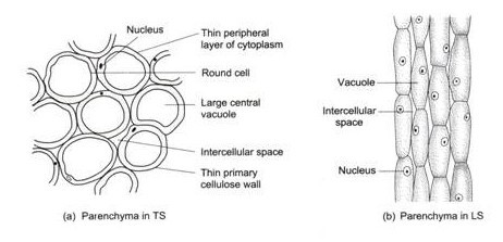
Parenchyma tissue (meristematic though permanent)
- Living
- Protoplasm present.
- Cell walls generally thin. Sometimes thick.
- Round, oval, polygonal, isodiametric etc.
- Equally expanded.
- Intercellular space present.
- Vegetative propagation (e.g. cutting) via plant parts is possible due to meristematic potential of parenchyma cells.
Occurrence
- Major parts of plant body.
- Epidermis
- Cortex
- Spongy parenchyma + palisade parenchyma (together form mesophyll tissue).
- Xylem, phloem.
- Fruit pulp (juicy vesicle of a citrus fruit), endosperm of seed.
- Pith
- Edible flesh of fruit.
Special types of parenchyma
- Aerenchyma
- With large intercellular space.
- In cortex of aquatic plants.
- Contain well developed air space.
- Chlorenchyma
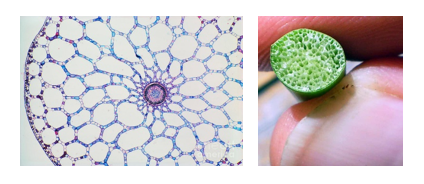
- Have chloroplast.
- They carry on photosynthesis.
- g. Spongy parenchyma, palisade parenchyma.
Function of parenchyma cells
- Main function is to store food in the form of starch, grain, fat etc.
- In case of xerophytic or succulent plants, they store water.
- Aerenchyma helps to float.
- Helps in exchange of gas.
- Chlorenchyma helps in photosynthesis.
Collenchyma tissue
- Living tissue.
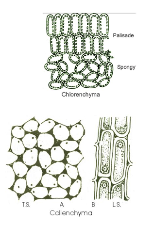
- Cell wall thick but not always.
- Thickness by cellulose and pectin.
- Uneven thickness.
- Elongated, rectangular, oblique cells with tapering or flat end.
- Water content is huge in cell wall. So the cells are extensible or elastic.
- Colouring agent: tannin, resin.
- Main source of mechanical support in the young stage of growing organs as they lack secondary tissue (Later on, secondary growth takes place. So, collenchyma cells are not required further).
Location
- Normally after epidermis.
- In growing organs.
- Mainly in supporting tissue of dicot leaf and stem.
- In periphery of stem and leaf.
- May be present in root cortex when roots are exposed to sunlight.
- Not found in root, stem and leaf of monocot.
- Mainly found in dicot.
Good to know
- The collenchyma cells have dynamic cell walls.
Sclerenchyma tissue
- Non-living.
- Cell lacks living protoplasm.
- Cell walls are thick because of lignin deposition. So, called lignified cell.
- Chloroplast absent.
- Also called mechanical tissue.
Functions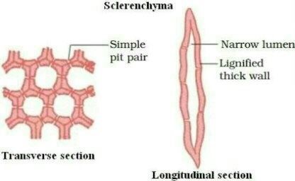
- Provides mechanical support i.e. strength to the cell.
Pit
- Pit is a characteristic of dead cell.
- It is caused by unequal accumulation of lignin.
Classification of Sclerenchyma Tissue
- Fiber cell
- Sclereid cell
Fiber cell
- Elongated cells.
- With pointed tip***.
- Cell wall is always thick. Thickness is caused by lignin.
- Sometimes, the cell walls are so thickened that their cavities are reduced in size.
- In young stage, protoplasm is present. On maturation, protoplasm disappears. Cells become empty and dead. In rare cases, protoplasm may remain even upon maturity.
- Small size rounded pits are present.
- Cell shape is hexagonal in T.S.
- Cavities take upon pink colour when safranin is applied.
Occurrence of fibre
- Cortex, pericycle of stem (not in epidermis).
- In vascular tissue (in xylem and phloem).
- Can also be present as a single cell, continuous band.
Average length
- 1 to 3 mm in angiospermic plants.
- Exceptionally lengthy fibres: 20-550 mm.
- (Have economic importance)
Linum usitatissimum– Flax- Tisi- Linaceae.
Cannabis sativa– Hemp- Cannabaceae.
Corchorus capsularis– Jute- Paat- Malvaceae.
Boehmeria nivea– Ramie- Urticaceae.
On the basis of location of fibre
- Xylary fibre or wood fibre
- Extraxylary fibre or bast fiber
- Cortical fibre
- Phloem fibre
- Perivascular fibre
1. Xylary fibre or wood fibre
- Found in xylem tissue.
- Form the integrated part in xylem.
- Single celled, group or scattered arrangement.
- Develop from the same meristematic tissue like other xylem elements.
2. Extraxylary fiber or bast fiber
- Outside the xylem.
- Lengthy
- Pits are present.
- End wall may be blunt.
- Thickness may be caused by hemicellulose.
a. Cortical fibre
- Present in cortex.
b. Phloem fibre
- Originate from primary or secondary phloem.
c. Perivascular fibre
- Found in the peripheral region of the vascular cylinder.
- Position: Inside the cortex tissue but not in the phloem tissue.
(inside the cortex, outside the phloem)
Sclereid cells
- More or less can be elongated.
- Isodiametric too but not so much.
- Found in: Xerophytic and hydrophytic plants.
Function
- Causes thickness and hardness.
Classification
- Brachy sclereid or stone cell.
- Macro sclereid.
- Ostea sclereid.
- Astro sclereid.
1. Brachysclereid or stone cell
- Looks like pedicel present in epidermis layer of seed and fruit. Also found in leaf and stem and the cortex of xerophyte.
- Shape stone like.
- Short cell.
- Found in pith and cortex of stem.
- Found in fruit pulp.
2. Macroslereid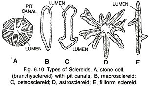
- Rod shaped.
- Found just beneath the epidermis.
- Widely distributed in leaf cell.
- In Sundarban.
- Also occur in seed coat.
- Commonly found in fruit, cortex and pith.
- May be present as a single cell or group.
- Found in cortex of leaf and stem of xerophytic plant.
3. Osteasclereid
- Bone shaped.
- Enlarged at both ends.
- Found in hypodermal layer after epidermis.
- Found in leaf, fruits and seeds.
4. Astrosclereid
- Star shaped with projection.
- Found in intercellular space of hydrophytic plant.
e.g. leaf cortex and stem cortex.

 Plantlet The Blogging Platform of Department of Botany, University of Dhaka
