One of the important fungi which influence human life belongs to the order Erysiphales. This fungi causes powdery mildew disease to plants, so named as the infected plant appear to be covered by a white, powdery material. This is none other than due to the powdery conidia, which are blown away easily. Their physiology and importance are discussed as follows-
Classification
Phylum: Ascomycota
Class: Ascomycetes
Order: Erysiphales
Family: Erysiphaceae
Genus: Erysiphe
Characteristics of Erysiphales
Their microscopic features are important in identification. they are –
- Mostly obligate parasite grow in host plants.
- Two types of haustoria present that is knob like and digitately branched.
- Both homothallic and heterothallic.
- Asexual reproduction by conidiophore.
- Sexual reproduction is followed by gametangial contact.
- Ascocarp is cleistothecium.
- Ascocarp contains various types of appendages.
- Mycelia conspicuous, growing mostly on plant surfaces.
- They are called powdery mildew fungi.
Best safe and secure cloud storage with password protection
Get Envato Elements, Prime Video, Hotstar and Netflix For Free
Best Money Earning Website 100$ Day
#1 Top ranking article submission website
Occurrence and importance
The members of the family Eryspipahceae cause a group of plant diseases commonly known as powdery mildews.
| Host | Causal species |
| Gooseberries | Sphaerotheca mors-uvae |
| Roses | Sphaerotheca pannosa |
| Apple | Podosphaera leucotricha |
| Cucurbits | Erysiphe cichoracearum |
| Cereal crops | Blumeria graminis |
| Ornamental plants | Erysiphe poligoni. |
Somatic structure
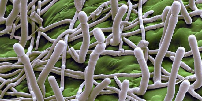
Hyphae: They are branched or unbranched, tend to grow closely appressed to the host surface. Some species produces aerial branches known as setae. Production of thick walled clamydospores are recognized in some species. They are thin walled, septate with each cell usually containing a single prominent nucleus.
Appresoria: Hyphae are anchored to the host by appressoria. They are simple, lobed structures or more complicated multilobed structure. They are produced in sequence along the length of hypha or in alternate or opposite fashion.
Haustoria: several types are recognized based on the light microscopic studies of few species of Erisiphales. They appear to form slender neck region and the body which may be globose, pyriform or lobed. Haustorium is seperated from the cytoplasm of the host cell by by a so called extrahaustorial matrix and extrahaustorial membrane.
Asexual reproduction
The chief source of dissemination of this fungi is conidia, produced in enormous number on vertical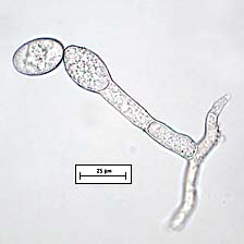 conidiophores. They are hyaline and erect. Most of the species contains conidiophore which are unbranched (except Leveillula).The basal cell is referred to as foot cell while the terminal cell is called either generative cell or conidiogenous cell.
Conidia develop singly or in chains. They remain firmly attached end to end with mature conidia at the distal end of the chain and immature conidia at the proximal end (basipetal development). They are one celled and appear hyaline when viewed with light microscopy. Each is thin walled, uninucleate and highly vacuolate. Structure known as fibrosin body is noted.
Sexual reproduction
Sexual reproduction may occur in homo- or heterothallic. Uninucleate gametangia arise from two closely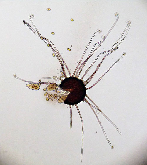 placed hyphae, which in the beginning are similar but later becomes differentiated. The antheridium is slender while ascogonium is curved and swollen. Both are uninucleate and separated from the hypha by a septum. Lower cell is called stalk cell.
The development of ascocarp is not yet resolved. There is a controversy as to where the sexual nuclei originates from. One school of thought regarded the two hyphal branches that coil around one another as functional gametangia with antheridial nucleus passing into the ascogonium rendering it binucleate. Another school of thought states it is the division of female nuclei into dikaryon and this dikaryon further divides to form 4 cells. They are separated into 3 cells with the middle having binuleate cell. At maturity of the cell, cleistothecium is developed, where the asci contains 1 to 8 ascospores. The external ennvironment induces the splitting of ascocarp thus spore is rereased.
Classification on the basis of cleistothecial appendages
There are four kinds of appendages present in a cleistothecium.
- Appendages flaccid, myceloid type.
- Appendages rigid, dichotomously branched at the tip of the apex.
- Appendages rigid, tip circinately coiled and hook like appendages present at the tip of the apex.
- Appendages rigid, spear like, arising from a bulbous base and tip pointed.
(a)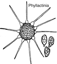 (b)
(b)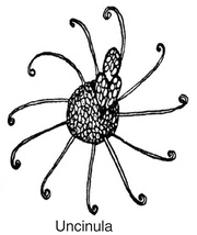 (c)
(c)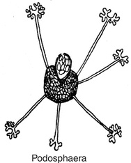 (d)
(d)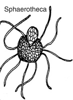
fig: (a) bulbous appendage, (b) circinoid appendage, (c) Dichotomous appendage, (d) myceloid appendage
References
- Introductory Mycology (C. J. Alexopoulos; C.W. Mims; M. Blackwell )
- An introduction to Fungi 3rd Edition (H C Dube)
 Plantlet The Blogging Platform of Department of Botany, University of Dhaka
