When we observe a living cell, we can see a variety of living bodies of definite structures and functions suspended in the cytoplasm, known as organoids or organelles. These organelles are the main sites for various cytoplasmic activities. Some of these are concerned with the chemical activities or metabolism of the cytoplasm. The ribosome is one of them. Ribosomes are particles composed of ribonucleic acid (RNA) and proteins. These particles are an integral part of the rough endoplasmic reticulum and microsomes. But they also exist freely in the cytoplasm, e.g. in young meristematic cells.
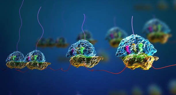
Ribosomes are often attached in rows or clusters. These clusters or strings of ribosomes are called polyribosomes or polysomes. They consist of ribosomes that are very active in protein synthesis and are held together by a string of messenger RNA (mRNA).
Best safe and secure cloud storage with password protection
Get Envato Elements, Prime Video, Hotstar and Netflix For Free
Best Money Earning Website 100$ Day
#1 Top ranking article submission website
Discovery
- Hanstien and Altmann (1882) found some very small granules in the cytoplasm of cells. They termed this granule as microsome.
- Claude (1954) got similar types of particles after centrifuging the liver cells and termed these particles as microsomes like Hanstien and Altmann.
- In 1955, George Emil Palade discovered ribosomes and described them as small particles in the cytoplasm that mainly remain with the endoplasmic reticulum. He, along with other scientists, discovered that ribosomes performed protein synthesis. For his work, Palade won the Noble prize in 1974.
- Later, Richard B. Roberts first proposed the term “ribosome” at the end of the 1950s.
- Sevetz (1965) observed that kind of particle under an electron microscope and described microsome as a small portion of rough walled endoplasmic reticulum. It consists of –
- the membrane of E.R.
- many small particles i.e. ribosome
Distribution
- Generally, they are found on the rough-walled endoplasmic reticulum.
- In the case of a eukaryotic organism, ribosomes are found within the mitochondria, chloroplast, and also in the cytoplasm freely.
- In the case of prokaryotic organisms, ribosomes are found in the cytoplasm freely.
- The ribosome granules in young meristematic cells lie freely in the cytoplasmic matrix.
Classification based on S-value
Swedish biochemist Svedberg observed that after keeping ribosomes in less concentration of Mg (0.001M) solution if it is centrifuged then the ribosomes are sedimented. The sedimentation rate is constant for similar types of ribosomes and it is called as “Svedberg constant” or “S-value”.
Based on S-value, ribosomes are two types:
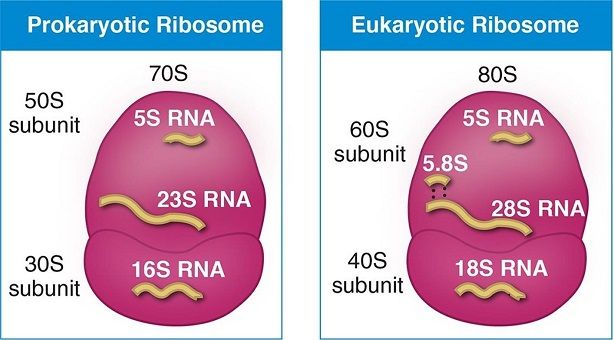
- The 70S ribosome (found in prokaryotic organisms like bacteria, blue-green algae, etc.)
- The 80S ribosome(found in eukaryotic organisms)
Each ribosome consists of two subunits: a smaller sub-unit and a larger sub-unit. Both subunits are composed of long strands of RNA.
70S ribosome particles split into 50S larger subunit and 30S smaller sub-units. Actually, these two sub-units occur freely in bacteria cytoplasm and unite only during protein synthesis. Similarly, 80S particles split into 60S and 40S subunits. The 40S sub-unit occurs above the 60S subunit forming a cap-like structure. The 60S subunit is dome-shaped and attaches to the membranes of canaliculae in fixed ribosomes.
The association or dissociation of the two sub-units depends on Mg2+ ion concentration. Low Mg2+ concentration causes separation of two sub-units, while high Mg2+ concentration causes association of them into the complete ribosome. Moreover, at a high concentration of Mg2+ ion in the cytoplasmic matrix, two or more ribosomes become associated with one another to form dimer or polymer.
Chemical Composition
D. Watson (1958) isolated ribosome and analyzed from E. coli. He observed that there are two main components:
- 40% RNA (specially rRNA)
- 60% Protein
- In E. coli, the ribosome is about 22% of their total dry weight.
- rRNA is about 98% of the total RNA.
Spirin (1964) has shown that each of the two ribosomal sub-units has the form of a long RNA-protein filament coiling to produce the globular form of the mature ribosome. The larger subunit has one large RNA molecule as well as a small RNA molecule, along with several protein chains. On the other hand, the smaller sub-unit consists of just one RNA molecule and several protein chains.
Physical Structure
- At first, people believed that the ribosome has a uniform structure. Nauringa (1967) proved that it has two components. The larger one ( 50S subunit of 70S ribosome) is a pentagonal structure of 100Å that attaches with a globular structure of 40-60Å in diameter.
- In bacteria and other prokaryotes, chloroplast, and mitochondria of eukaryotes, ribosomes are 150Å in diameter. However, ribosomes in the cytoplasm of plant and animal cells are about 140 to 200Å in diameter.
- The ribosome is bounded by a membrane (thickness 70Å) that doesn’t dissolve by ribonuclease, however, dissolve with protease. It means, the wall is made up of protein. There are many soluble protein present in ribosome which are basically different enzymes.
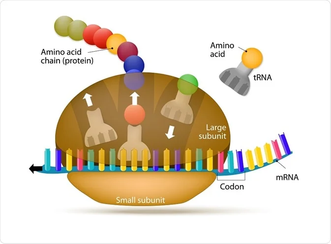
- Ribosomes have a groove at the junction of large and small subunits.
- The m-RNA remains in the gap between both ribosomal subunits, where the ribosome protects a stretch of some 25 nucleotides of mRNA from degradation by ribonuclease.
- From this groove, a canal or tunnel extends through the large sub-unit and opens into the lumen of the endoplasmic reticulum.
- The groove between the two ribosomal subunits is the site for the synthesis of polypeptides. After that, they pass through the tunnel of the large subunit into the endoplasmic reticulum.
Functions of Ribosome
Ribosomes are the protein factories of cell. The one & only function of them is to synthesize protein by the process of Translation.
Attached Ribosomes: Attached to the endoplasmic reticulum and other organelles.
- These ribosomes provide space and enzymes for the synthesis of proteins in the cell. The ribosomes bound to the ER membranes synthesize:
- integral proteins for cellular membranes
- lysosomal proteins and
- secretary proteins for export as secretions.
Free Ribosomes: Found freely in cytoplasm.
- The free ribosomes produce structural and enzymatic proteins for use in the cell itself.
References & Other Links
- Class lecture of Chandan Kumar Dash Sir (Lecturer, Dept. of Botany, University of Dhaka).
- Book- “Cytogenetics, Evolution, Biostatistics & Plant Breeding” by R.S. Shukla, P.S. Chandel.
- Ribosome from Scitable by Nature Education .
- Ribosome from Slideshare.

very informative article indeed. If possible please do add some more functions of ribosome like it can cause extrachromosomal disorder in organisms and anything you find new.
Thank you so much for your feedback. I’ll try to find and add some more information regarding this part.
Very much informative and impressive💙. Thanks for the infos.
Thanks for your appreciation.
Your article helped me a lot, is there any more related content? Thanks!
Awesome! Its genuinely remarkable post, I have got much clear idea regarding from this post
This is really interesting, You’re a very skilled blogger. I’ve joined your feed and look forward to seeking more of your magnificent post. Also, I’ve shared your site in my social networks!
I do not even understand how I ended up here, but I assumed this publish used to be great
For the reason that the admin of this site is working, no uncertainty very quickly it will be renowned, due to its quality contents.
Let’s hope for the best✌
For the reason that the admin of this site is working, no uncertainty very quickly it will be renowned, due to its quality contents.
This is really interesting, You’re a very skilled blogger. I’ve joined your feed and look forward to seeking more of your magnificent post. Also, I’ve shared your site in my social networks!
Thanks for the appreciation. Means a lot.
This is really interesting, You’re a very skilled blogger. I’ve joined your feed and look forward to seeking more of your magnificent post. Also, I’ve shared your site in my social networks!