
Meristematic tissue is the plant tissue that has the ability to divide actively throughout its life. It consists of undifferentiated cells capable of cell division.
Apical Meristamatic tissue is one kind of meristem which stays at the tip of a plant shoot or root and causes the shoot or root to increase in length. There are Different Theories regarding the Structural Development and Differentiation of Apical Meristem.
In this article, Classification of Meristematic tissue and the Theoris of Apical Meristem will be discussed.
Best safe and secure cloud storage with password protection
Get Envato Elements, Prime Video, Hotstar and Netflix For Free
Best Money Earning Website 100$ Day
#1 Top ranking article submission website
Classification of Meristematic Tissue
Meristematic tissue can be classified in four ways:
1. Origin
• Primary
• Secondary
2. Function
- Protoderm
- Pro-cambium
- Ground
3. Position in plant body
• Apical
• Lateral
• intercalary
4. Stage of development
• Pro
• Primordial
Origin
Primary Meristem
- Builds up the primary part of the plant (in apex of root, stem, leaf).
- Consists of Promeristem.
Secondary Meristem
- Appears at later stage of development of plant body organ.
- Always arises from permanent tissue.
- Are found at lateral side of stem and root.
Function
Primary meristem at the apex of stem and root is distinguished into three meristematic tissue:
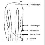
Protoderm
• Outermost tissue which develops into epidermis.
-
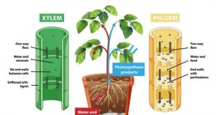
Procambium
• Develops into primary vascular tissue.
-
Fundamental or Ground
• Develops into ground tissue and pith.
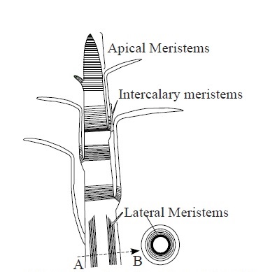
Position in the Plant Body
-
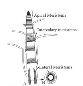
Apical Meristem
• Lies at the apex of stem and root of vascular plants.
• Increases organ length.
• Initiation of growth takes place by one or more apical cells situated at the tip of
organ. -
Lateral Meristem
• Composed of initials which divide mainly in one plant (periclinally).
• Increases organ diameter.
• Example: Cambium, cork cambium. -
Intercalary Meristem
• Portion of apical meristem that becomes separated from apex during
development by permanent tissue.
• Internodal in position.
Stage of Development
- Promeristem or Primordial meristem: Promeristem is the region of new growth in a plant body where the foundation of new organ or parts of organ is initiated.
Theories of Structural
Development and Differentiation of
Apical Meristem
There are Three theoeries of Structutal Development and Differentiation of Apical Meristem. These are:-
-
Apical cell theory
• Applicable for both shoot and root.
-
Histogen theory
• Applicable for both shoot and root.
-
Tunica-corpus theory
• Applicable only for shoot.
Apical Cell Theory
• Proposed by Nageli in 1858.
• Solitary apical cells occur in many of Algae, bryophytes and vascular cryptogams.
• Applicable for cryptogams.
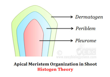
Histogen Cell Theory
• Proposed by Hanstein in 1870.
• Primordial meristem was sharply separated into three distinct zones of histogens
(Dermatogen, Periblem, Plerome).
• Each zone consists of a group of initials called tissue builders.
These three zones are described below:
1. Dermatogen
-
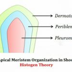
Histogen Theory. Source Single outer most layer of the cell of
apical meristem which later gives rise
to epidermis of stem and root. - Single layered in root but merges into
Periblem at the apex. - Outside Periblem, forms many new
cells resulting into a small celled tissue
(Calyptrogen). - Calyptrogen gives rise to root cap.
2. Periblem
• Found internal to dermatogen.
• Middle region of apical meristem.
• Single layer at the apex but becomes multilayer in the center (for root and stem).
• Develop into cortex.
3. Plerome
• Central meristematic region of stem and root apex.
• Develops and differentiate into central stele (vascular tissue, ground tissue, pericycle, medullary ray, medulla).
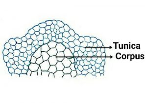
3. Tunica Corpus Theory
• Proposed by Schmidt in 1924.
• According to the theory, there are two
zones of tissue in Apical meristem.
i. Tunica
ii. Corpus
• No difference between tunica and corpus layer in primitive types of plants
(Lycopodium, Selaginella).
• Shoot apex has
i. Weak tunica corpus layer in Pinus plants.
ii. Clear tunica corpus layer in Angiospermic plants.
i. Tunica
- Peripheral layer consisting of one or more peripheral layers of cells.
- Shows anticlinal division resulting in surface growth.
- Each layer of tunica arises from a group of separate initials.
ii. Corpus
- A mass of cells enclosed by the tunica.
- Cells are large.
- Mas grows in volume as arrangement and plane of division are irregular.
- Corpus has one layer of initial.
Reference:
1. Class Lecture of Parveen Rashid, PhD
Professor, Department of Botany, University of Dhaka.

I don’t think the title of your article matches the content lol. Just kidding, mainly because I had some doubts after reading the article.