All living things, as well as human beings, are made up of cells. Some organisms have only one cell during their whole lifespan by which they carry out all the physiological processes they require to survive. These are unicellular organisms. For instance, bacteria, yeast, etc. In contrast, there are also many other organisms whose bodies are made up of two or more cells. Thus, these are called multi-cellular organisms. For instance, higher plants, animals, human beings, etc. Multi-cellular organisms grow up and reproduce by dividing their cells.
Cell Division
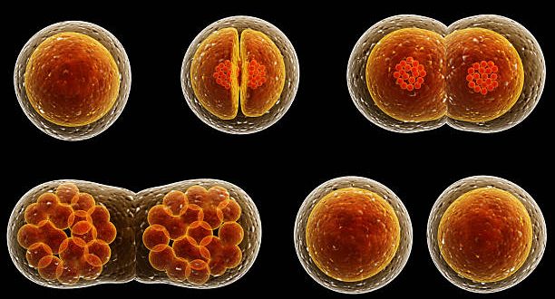
Cell division is the basic process by which a parent cell divides into two or more daughter cells. However, it occurs as a small part of a cell cycle. Both prokaryotic & eukaryotic organisms grow up and reproduce by this process.
There are three stages of cell division:
- DNA replication
- Nuclear division (karyokinesis)
- Cytoplasmic division (cytokinesis)
Best safe and secure cloud storage with password protection
Get Envato Elements, Prime Video, Hotstar and Netflix For Free
Best Money Earning Website 100$ Day
#1 Top ranking article submission website
Also, 3 types of cell divisions occur in living organisms:
- Amitosis
- Mitosis and
- Meiosis
Discovery of Cell Division
| Year | Scientist (s) | Contribution |
| 1879 | Dr. Schleicher | First gave the word ‘Karyokinesis’. Here, ‘karyo’ = nucleus & ‘kinesis’ = division. |
| 1882 | Walter Flemming; German biologist and a founder of cytogenetics |
Observed, along with nucleus, some thread-like structures were also divided equally and name the incident ‘Mitosis’ (mito =thread, sis= division). |
| 1888 | Waldeyer | Observed that the thread-like structure took a certain stain. He named the thread-like structures ‘chromosomes’. Here, chroma = color, soma = absorb. |
| T. O. Caspersson | Said that DNA replication occurs in Prophase. | |
| 1925 | Polyster et. al | Through ‘Quantitative Feulgen Micro-spectrophotometry DNA Stain method’, they observed that if the light is being passed through interphase, the cot values (described below) were
Interphase – 4C Prophase – 4C Metaphase – 4C Anaphase – 2C Telophase – 2C Cell – 2C |
Conclusion
In conclusion, in the same species, the amount of DNA is-
- 2C in diploid somatic cell.
- C in the gametic cell.
- In zygote is 2C.
- In interphase is 4C.
- It becomes 2C in the mitotic anaphase.
Therefore, DNA replication or chromosome duplication occurs in interphase stage, not in prophase.
Cot Value: A technique for measuring the complexity (i.e size) of DNA or genome. Co = concentration of DNA and t = time taken for renaturation. Low cot value indicates more no. of repetitive sequences. On the other hand, a high cot value shows more no. of unique sequences or less no. of repetitive sequences.
Cell Cycle
Cell cycle is the series of events that take place in a cell when it grows and divides. Howard & Pelc gave the cell cycle during 1953. They used the [3H] thymidine isotope for this reason. Subsequently, they observed 4 steps in the cell cycle:
- Non-function
- Replication of DNA
- Non- function
- Division
In short, all these 4 steps are divided into 2 major stages:
- A long, undividing stage or, interphase or, I-phase (Non-function, Replication & Non-function)
- A short, dividing stage or, mitotic phase or, M-phase (Division)
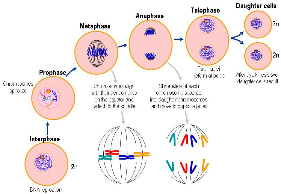
Mitotic phase is further divided into 5 sub-stages:
- Prophase (pro = initial)
- Pro-metaphase (pro= initial, meta = middle)
- Metaphase (meta = middle)
- Anaphase (ana = move)
- Telophase (telo = end)
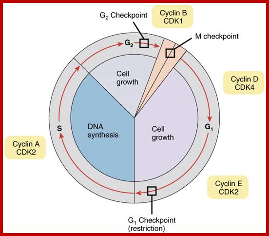
G1– Checkpoint
Checks for:
- Size of cell
- Nutrients
- Growth regulator
- DNA damage
G2– Checkpoint
Checks for:
- DNA replication completeness
- DNA damage
M- Checkpoint
This checkpoint is also known as the ‘spindle checkpoint’.
Checks for:
- Chromosome attachment to the spindle.
If the DNA is beyond repair the checkpoint mechanism can transmit a signal that leads to-
- The death of the cell or,
- A permanent cell-cycle arrest (known as senescence).
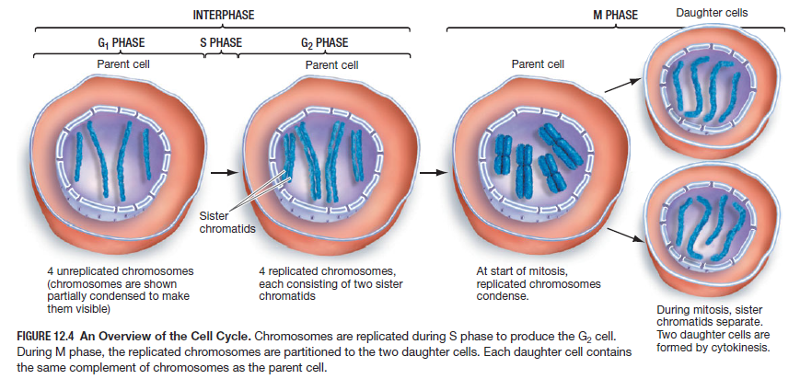
Interphase or I-phase
The time between the end of the telophase (end of forming new cells) and the beginning of the next M-phase is called interphase or I-phase. Subsequently, we can say, interphase is the preparation stage before cell division.
- It lasts about 10-30 hours depending on different conditions.
- During this phase, the cell grows by synthesizing biological molecules such as carbohydrates, lipids, proteins, nucleic acids, etc.
Moreover, Interphase has 3 sub-phases:
- G1-phase
- S-phase
- G2-phase
G1-phase
The gap between the end of telophase and the beginning of DNA synthesis is the G1 phase. Things that happen during this phase are as follows:
- Initial growth of newly formed cells.
- Various biological molecules are synthesized.
- Most importantly, takes preparation for DNA replication.
Nevertheless, if extracellular conditions are unfavorable, the G1 phase delays. Then the cell enters into a specialized resting state termed G0(G zero). However, in this state, the cell can remain for days, weeks, or even years. Many cells can stay in this state until the cell or the organism dies.
S-phase
The part of the interphase where DNA is synthesized is S-phase.
- Each chromosome is duplicated by DNA replication.
- Formation of new nucleosome.
- Lasts for about 6 to 8 hours.
G2-phase
- Cytoplasmic organelles for instance centrioles, mitochondria, golgi body, chloroplast, etc are doubled.
- Formation of proteins for spindle aster synthesis.
- Lasts for about 2 to 5 hours.
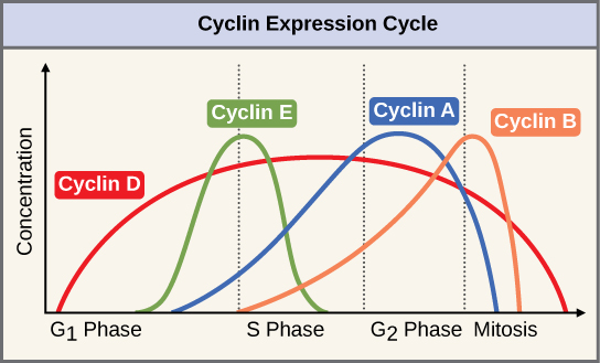
Central components of the cell-cycle control system are members of a family of protein kinases. They are known as cyclin-dependent kinases or Cdks. The foremost vital of these Cdk regulators are proteins known as cyclin. There are four classes of cyclin classified by the cell cycle stage at which they bind Cdks and function. Three of these classes specifically work at three checkpoints of a cell cycle but one doesn’t.
| Cyclin-Cdk Complex | Cyclin | Checkpoint | Function |
| G1/S Cdk | Cyclin E | G1 Checkpoint | Activate Cdks in late G1 and thereby help trigger progression through Start resulting in a commitment to cell cycle entry. Their levels fall in S-phase. |
| S Cdk | Cyclin A | G2 Checkpoint | Bind Cdks soon after progression through Start and help stimulate chromosome duplication. S-cyclin levels remain elevated until mitosis and this cycle also contributes to the control of some early mitotic events. |
| M Cdk | Cyclin B | M Checkpoint | Activate Cdks that stimulate entry into mitosis at the G2/M transition. M-cyclin levels fall in mid-mitosis. |
| G1 Cdk | Cyclin D | _ | Helps govern the activities of the G1/S-cyclins, which control progression through Start in late G1. |
Mitotic phase or M-phase
- During this phase, all the chromosomes divide into two daughter cells.
- Many structural and physiological changes take place.
- Chromatins are packed into chromosomes.
- The disappearance of the nucleolus and nuclear membrane also occurs.
- Endoplasmic reticulum and Golgi bodies get fragmented into small vesicles and prevent movement.
- Microtubules assemble into spindle fibers.
- Actin filament (a kind of protein ) forms a contractile ring for the cytoplasmic division.
References
- Class lecture of Chandan Kumar Dash Sir (Lecturer, Department of Botany, University of Dhaka)
- Cell & Molecular Biology Concepts and Experiments by G. Karp
- Molecular Biology of The Cell by Bruce Alberts.
Revised By
- Khaleda Akter Shompa on 30 July 2021.
- Tarannum Ahsan on 2nd August 2021.
- Tarannum Ahsan on 9th August 2021.
 Plantlet The Blogging Platform of Department of Botany, University of Dhaka


This was easy and helpful.
Thank you so much
Checkpoint discussion was really clear and informative.
Thank you. Let me know if you need to add anything else here.
Very much reader friendly & well organized article. Thanks for sharing tricky studies in such an easy way.
Thanks a lot. Hope this is helpful for you..
I was looking for some explanation of cyclin and CDK protein function but the article was very informative and easy to read. Keep up the good work.
I’ll try to add more information about it. Thanks for letting me know.