
Secondary growth is characterized by an increase in thickness or girth of the plant. It is caused by cell division in the lateral meristem. Herbaceous plants mostly undergo primary growth, with little secondary growth or increase in thickness.
Secondary growth is initiated by the activity of the vascular cambium as far as the steler region is concerned. This intrafascicular cambium is absent in the open vascular bundles of the monocot stem, thus the process cannot take place in Monocot plants.
In this article, Secondary Growth in Dicot Stem and Root will be discussed briefly.
Best safe and secure cloud storage with password protection
Get Envato Elements, Prime Video, Hotstar and Netflix For Free
Best Money Earning Website 100$ Day
#1 Top ranking article submission website
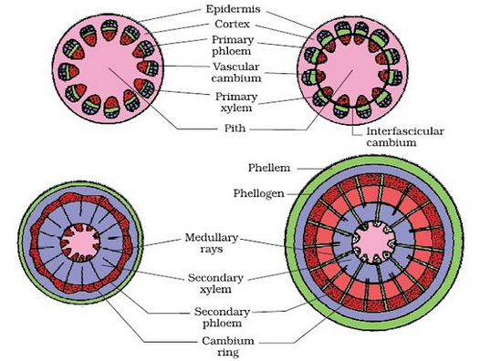
Secondary Growth in Dicot Stem
Because of the presence of Cambium, Secondary Growth takes place in Dicot plants. Secondary Growth of Dicot Stem is presented below:
- In perennial dicotyledons (shrubs and trees) after the primary tissues are fully
formed, the cambium becomes active and begins to cut off new secondary
tissues in the stellar region. - Another strip of meristem, the cork cambium makes it appearance in the peripheral region and begins to form other secondary tissues in that stellar region.
- All the secondary tissues are added to the primary ones and as a result the stem increases in thickness.
- The increase in thickness, due to the formation of new secondary tissue by the activity of cambium and cork cambium in the stellar and extra-stellar regions, are called secondary growth.
Activity of Cambium
Cambium Ring
Some of the medullary ray cell, mostly in a line with the fascicular cambium becomes meristematic and form a strip of interfascicular cambium. The joins on to the fascicular cambium on either side and forms a complete ring known as the cambium ring.
Secondary Tissue
- As a whole, the cambium ring becomes actively meristematic and gives off new cells both externally and internally. Those cells are cut off on the outer side are gradually modified into the elements of the phloem. These contribute the secondary phloem. Those cells are cut off on the inner side are gradually modified into various elements of xylem and form secondary xylem.
- The cambium is more active on the inner side than on the outer.
- Continuous formation of secondary xylem and the pressure exerted by it, the cambium, phloem and surrounding tissues are gradually pushed outwards and for the same reason some of the primary tissues get crushed. The primary xylem remains more or less intact in the center.
- Here and there the cambium forms some narrow bands of parenchyma, radially elongated, passing through the secondary xylem and secondary phloem. These are the secondary medullary rays.
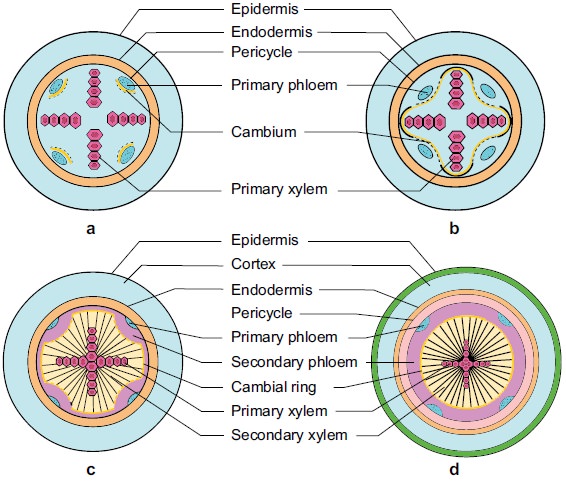
Secondary Growth in Dicot Root
Secondary Growth in Dicot Root takes place by the Formation of Cambium and development of secondary tissue. These are described as follows:
- On the initial stage of secondary growth, a few parenchymatous cell beneath each group of phloem becomes meristematic and thus as many cambial strips are formed as the number of phloem groups. The cambial cells divide tangentially again to produce secondary tissue.
- Some of the cells of the single layered pericycle become meristematic lying against the protoxylem groups which divide and form few layers of cells. The first formed cambium now extends towards both of its edges and reaches the innermost derivatives of the pericycle, thus giving rise to a complete ring of cambium.
- The cambium ring passes internal to phloem and external to xylem. So it becomes wavy in outline. The cambial cells produce more xylem element than phloem.
- The first formed cambium produces secondary xylem much earlier and the wavy cambium ring ultimately becomes circular. Now whole of the cambium ring becomes actively meristematic and behaves as to stem giving rise to secondary xylem on its inside and secondary phloem on its ouformed.
- The secondary vascular tissues form a continuous cylinder and usually the primary xylem get embedded on it. At this stage distinction can be made only of exarch primary xylem located in the center. The primary phloem elements are generally seen in crushed condition.
- The cambial cells that originate from the pericycle lying against the groups of protoxylem function as ray initials.
Periderm
- Simultaneously the periderm develops lenticel in the outer region of the root. The single layered pericycle becomes meristematic and divides and giving rise to cork cambium or phellogen.
- It produces a few brownish layers of cells or phellogen towards the outer side and phelloderm on the inside. The phellogen does not contain chloroplasts. The pressure caused by the secondary tissues ruptures the cortex with endodermis which is ultimately sloughed off . The epiblema dies out earlier. Lenticel may also be formed.
Reference:
1. Class Lecture of Parveen Rashid, PhD
Professor, Department of Botany, University of Dhaka.
