In the kingdom Fungi, there are two major phyla- Ascomycota and Basidiomycota. These two together form a subkingdom of fungi named as Dikarya. They together were named such as members of both phyla maintain most part of their life cycle in dikaryotic stage meaning each cell contains two genetically different nuclei.
Related articles
Ascomycetes
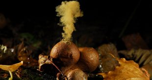
The members of Ascomycota are called Ascomyecetes. Fungi of Ascomycetes produce sac like structure called Ascus (pl. asci) in their life cycle. From that perspective, they are commonly known as the ‘Sac Fungi‘. Inside the ascus, typically 4-8 ascospores are produced which upon liberation and germination produces new ascomycetous fungi.
Best safe and secure cloud storage with password protection
Get Envato Elements, Prime Video, Hotstar and Netflix For Free
Best Money Earning Website 100$ Day
#1 Top ranking article submission website
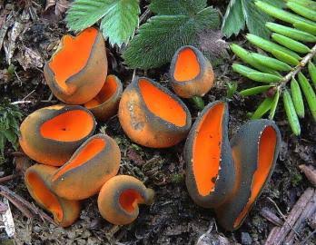
As Ascomyectes are huge in number, it seems to be so tough to identify all of them. However, scientists follow some methods to identify the members of sac fungi.
In this article, few popular methods of identifying Ascomyectes have been discussed.
Identification of Sac Fungi
There are two basic ways to identify Ascomycetes.
- By observing their unique structure.
- From complex test.
1. By Observing Unique Structure
This method of identification doesn’t claim complicated test or comparative studies. Only the pure and ensured observance is required. So, it is much performed in the laboratories by both students and teachers.
As already discussed, the primary morphological feature that distinguishes the members of Ascomycetes from all other fungi is ‘Ascus’. Ascus ( pl. Asci ) is a sac like structure containing the ascopores.

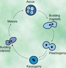
Ascus is generally cleaved from mycelium by free cell formation after karyogamy and meiosis (see the figure below). There are 8 ascospores formed within each ascus but the number may vary from species to species.
Development of Asci
In life cycle, Ascomycetes have two distinct reproductive phases:
- Sexual phase: Involving in the formation of the asci and ascospore/s .
- Asexual phase: Involving in spore production.
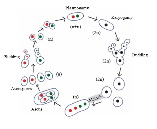
In sexual phase, during plasmogamy and karyogamy, two compatible nuclei are brought together in the same cell and fuse to produce ascus later. The fusion of two compatible nuclei can occur in variable ways such as:
- Two morphologically similar gametangia touch at their tips and fuse. The fusion cell develops into ascus.
- Two morphologically different gametangia (antheridia and ascogonia) are formed in this case. The male nucleus from the antheridium passes to the fusion point between antheridium and ascogonium and produce ascus. *Fusion cell is not developed here .
In this case, a single detached male cell works alike sperm (that’s why this process is called ‘spermatization’). The male cell becomes attached to the female receptacle. Through septal pores, the nuclei of male cell migrate to the ascogonium.
However the karyogamy occurs, the resultant zygotes later develops into an ascus. The ascus may be spherical to elongated, cylindrical, ovoid or globose (means round) shape. They may be arranged in scattered fashion or in a stalk or hymenium.
2. From Complex Test
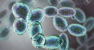
Yeast fungi are generally included in the phylum Ascomycota. But members of several other groups of fungi also show yeast stage in their life cycle. So, it seems to be a hard task to determine whether a fungi is ascomyctous or not simply by observing its yeast stage.
In that case few methods are applied to identify or determine Ascomycetes fungi. Some of such methods are following:
a) Diazonium Blue B (DBB) staining method
Others fungi except Ascomycetes shows positive result in DBB staining. Where the Ascomycetes are primarily nonstaining. So under microscope, it’s a simple task to determine whether it is Ascomycete or not.
b) Transmission electron microscopy (TEM)
The cell wall of Ascomycetes are of two layers- a thick, transparent inner and a thin, dense outer layer (in TEM). This scenerio is different from basidiomycetes or others. So it helps in determining Ascomycetes.

c) Observing Various Formation
In yeasts the manner in which new walls form in budding usually can be used to distinguish the Ascomycetes. In buds of ascomycetous yeasts, new wall layers are continuous with the parent wall. This feature is different than basidiomycetous buds. Thus ascomycetous fungi can be identified observing the bud wall.
Much more practice and basic knowledge about Ascomycetes is required for the exact identification of Ascomycetes.
Written by
Tarek Siddiki Taki
 Plantlet The Blogging Platform of Department of Botany, University of Dhaka

The article is short but precise in information. Overall, it is nice.
Please, do consider…
1. adding real photo of ascus.
2. adding photos of the three types of sexual ascus development.
N.B: Do mention the source of the image in the caption box.