The cell wall is structurally and chemically different from the protoplasm. The growth in thickness of walls is evident in both primary and secondary walls. The initiation of the cell wall and how it grows is being studied here.
Origin of the Cell wall
The wall formation is first evident at the close of nuclear division. In higher plants, cell wall biosynthesis begins at the center of two daughter nuclei after the cytokinesis phase at the end of the telophase stage. Protoplasmic substances like pectin are deposited in the form of droplets in the equatorial region between two daughter nuclei. These pectin droplets gradually increase in size and unite with each other and form the fluid plate. So the partition formed between two protoplasms is known as cell plate.
When the cell plate appears in the median part of the equatorial plane of the phragmoplast, the fibers of the phragmoplast disappear in this position. The cell plate occupies the former position of the phragmosome.
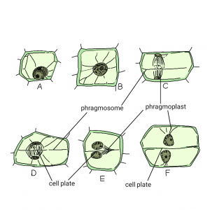
Figure- Origin of cell wall
Description of the above figure is as follows:
Best safe and secure cloud storage with password protection
Get Envato Elements, Prime Video, Hotstar and Netflix For Free
Best Money Earning Website 100$ Day
#1 Top ranking article submission website
- A– cell in a non- dividing state.
- B– Nucleus in prophase and located in the middle of the cell.
- C– Nucleus in early anaphase; laterally mitotic spindle is connected to peripheral cytoplasm by a cytoplasmic layer, the phragmosome;
- D– Daughter nuclei in telophase, the barrel-shaped spindle between the nuclei is the phragmoplast; cell plate appears in its equatorial plane.
- E– Cell plate intersects one of the walls of the mother cell.
- F– cell division is completed and the cell plate occupies the former position of the phagosome.
Growth of the Cell wall
The growth of the cell wall involves the growth in the surface area as well as in thickness. New cell wall materials like matrix polysaccharides and cellulose microfibrils are deposited over the expanding cell wall. the cellulose microfibrils are formed between the plasmalemma and the outer envelope.
The cell wall is thin, soft, elastic in nature. The growth of cell walls occurs due to the expansion and deposition of new materials. The cell wall is thin in immature cell and gradually increase in thickness towards maturity.
There are two theories regarding the increase of thickness of cell wall. These are as given below:
Apposition
The growth of the wall through the deposit of new layers from inside (from the cytoplasm) is called apposition. In apposition, new wall layers are laid and growth of wall in new cells occurs. Closely associated microfibrils are incorporated on the surface of present microfibrils and form a new layer.
Intussusception
The expansion of the cell wall in plants as a result of the introduction of new molecules of cellulose and protection into the already formed wall is called intussusception. Intussusception was previously contrasted to apposition. Intussusception is growth by deposition of closely associated new microfibrils between existing microfibrils of cell walls. The growth by apposition is usually centripetal.
References & Other Links
- Class Lecture of Parveen Rashid, PhD (Professor, Department of Botany, University of Dhaka).
- Book: Plant Anatomy by B.P.Panday (page-75-76)
- Picture- picture from Plant Anatomy- B.P.Panday (page-76) edited.
Revised by
- Saifun Nahar Smriti on 21 February, 2021.
- Khaleda Akter Shompa on 28 July, 2021.
 Plantlet The Blogging Platform of Department of Botany, University of Dhaka
