Psilotum is commonly known as Whisk-fern.
Salient features of Pilotum
- The sporophytes are dichotomously branched with an underground rhizome and upright branches.
- The upright branches are leafless.
- Rhizoids are present instead of root.
- Stem has a relatively simple vascular cylinder.
- The sporangia are born in groups (trilocular) and form synangia.
- The spores produced are all alike (homosporous).
- The development of gametophytes is exosporic and forms monoecious subterranean gametophyte.
- The development of the embryo is exoscopic.
Exasporic: The gametophyte is free-living and develops outside of the spore wall.
Exoscopic: Having the apex of the embryo pointed toward the neck of the archegonium.
Sporophyte
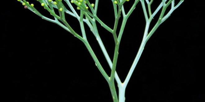
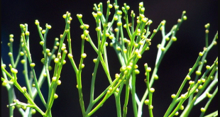
The plant body of Psilotum is a sporophytic branched rhizome system and dichotomously branched, slender upright green aerial system that bears small appendages and synangia.
Aerial stem
Any one of the rhizome tips may turn upward and undergo several dichotomies to give rise to a green aerial shoot.
The aerial shoots are slender, generally erect but maybe pendant in epiphytes (P. flaccidum). They are perennial and become shrubby, by repeated dichotomies, and sometimes obtain a height of up to one meter.
The aerial axis may be cylindrical at the base, furrowed on the upper parts, but somewhat flattened with these Longitudinal ridges at the top.
The basal part of the aerial axis is smooth but the distal part became small, scaly appendages and synangia.
The aerial stems are photosynthetic and the general appearance is xerophytic, although the plants grow in a very moist environment.
In T.S. the aerial system is covered by
- An epidermis.
- Extensive cortical areas.
- Single-layered endodermis and
- Stele.
The stele is siphonostelic in the basal part which becomes actinostelic in the younger branches.
The epidermis is single-layered in which the outer cell walls are heavily cutinized and covered by a layer of cuticle. The epidermis is broken regularly by stomata.
The cortex is divided into three regions.
The outer portion (Chlorenchymatus) under the epidermis consists of elongated, lobed, chlorophyllous cells with intercellular spaces. 2-5 layered thick with chloroplastids and starch grains.
The middle cortex is 4 to 5 layers of vertically elongated thick-walled sclerenchymatous cells without intercellular spaces, walls apparently become lignified in the lower portion. These tissues provide mechanical support to the plant.
The inner cortex (parenchymatous cells); these cells are without intercellular space but contain more starch grains. The cortical tissue is bounded inwardly by a single-layered endodermis.
The extreme base, the stem is protostelic (actino stelic). In the middle portion, the stele is siphonostelic as the center of the xylem is occupied by a patch of elongated sclerenchymatous cells (sclerotic pith)
Rhizome
It bears numerous rhizoids, instead of roots, which perform the function of absorption and anchorage.
In T.S. the rhizome reveals an outermost epidermis, cortex, endodermis, pericycle, and stele.
The epidermis is indistinct and gives rise to 2 celled rhizoids.
The cortex is extensive, parenchymatous, and differentiated into outer, middle, and inner layers.
The outer cortical layer is characterized by the presence of intracellular endophyte mycorrhizae. The cells of the middle cortex are large with starch grains while the cells of the inner cortex are often dark brown in color because of the presence of Phlobaphen (an oxidative product of tannins).
The stele is protostelic (haplostele or actinostele) and is surrounded by a well-defined endodermal layer.
Appendages
These are small-scale structures helically arranged on the upper part of the aerial system. Internally the appendages are composed of parenchymatous photosynthetic cells., bounded by a single-layered cutinized epidermis. There is no stomata in the appendage. There is a vascular trace in the appendages of P. nudum), although minute leaf traces originate from the stele in P. flaccidum which fade out in the cortex.
Reproduction in Psilotum
By spores
Spore-producing structure: At maturity, many of the dichotomously branched aerial shoots become fertile and produce trilocular sporangia known as synangia.
The synangium(mature) generally has a three-lobed structure and each lobe of the synangium corresponds to a sporangium.
Loculicidal dehiscence: The locules of the largest capsules split as the fruit opens and the septa remain intact.
Vegetative Reproduction in Psilotum
The sporophyte, as well as the gametophyte of Psilotum (e.g. P nudum), propagates vegetatively through the production of Gemma.
They are small, multicellular, and ovoid structures developing on the surface of the rhizome (in sporophytic plant body) or prothallus (in gametophyte).
After detachment from the patient body, the gemmae of sporophyte may germinate to form a subterranean shoot, while the gemmae of prothallus on germination, form a new prothallus.
(Pilotum is the only plant in the plant kingdom where the vascular tissues develop in the gametophytic generation)
Development of Sporangium
The mode of development of each sporangium of the synangium of P. nudum is of the eusporangiate type. The sporogenous tissue is produced from a repeated division of primary sporogenous cells in various places.
Gametophyte
The mature gametophyte shows a striking similarity with a piece of sporophyte rhizome. It grows as a sporophyte with an associated fungus. Spores germinate exosporically to form the gametophyte. The mature gametophytes are brown, cylindrical, subterranean, radially symmetrical, and usually dichotomously branched but may sometimes become irregular.
The surface of the gametophyte is covered by long unicellular brownish rhizoids. The gametophytes grow by means of the apical meristem. In T.S. the gametophyte reveals cutinized peripheral cells which enclose many layered thin-walled parenchymatous cells. Many of the internal parenchymatous cells are filled with hyphae by a symbiotic fungus. It has been observed that in the gametophyte of tetraploid P. nudum, the center is occupied by the xylem with annular scalariform and reticulate tracheids, surrounded by phloem and endodermis.
Thus, Psilotum is the only plant in the plant kingdom where the vascular tissue develops in the gametophytic generation.
The external resemblance of the sporophytic rhizome and gametophyte, coupled with the presence of vascular tissue in the gametophyte, supported the homologous theory on the origin of alternation of generation.
Development of Antheridium
The antheridium develops from a single superficial cell (antheridial initial) of the prothallus. The periclinal division of the superficial cell produces an outer jacket initial and inner primary androgonial cell.
The outer jacket initially undergoes repeated anticlinal division and forms a single-layered jacket. The inner primary androgonial divides into various planes and produces a mass of developing androgonial cells, the last generation of which are the androcytes.
All this stage, the antheridium projects above the surface of the prothallus as a minute protuberance. Each androcyte eventually becomes a spirally coiled, multi-flagellate antherozoid and escapes by the disintegration of the operculate cell.
Development of archegonium
The archegonium is also developed from a single superficial cell (archegonial initial) of the prothallus. The initial cell undergoes periclinal division to form an outer primary cover cell and an inner central cell. The anticlinal division is followed by periclinal division and the outer cover cells produce a long projecting neck arranged in 4 vertical rows of cells with 4-6 cells in each row.
The central cell divides transversely to form an upper primary neck canal cell and divides to form 2-neck canal nuclei and generally, this division is not accompanied by wall formation resulting in a binucleate neck canal cell. The primary venter cell divides transversely to produce a large egg and a small ventral canal cell,
Fertilization
At maturity, the cell wall of the lower tier of neck cells becomes thick-walled and cutinized. The apical tier, however, breaks away in presence of water and the mucilaginous contents of the neck cells are released. Thus, a free passage is formed for the entry of the antherozoid.
Fertilization is accomplished by the union of a multi-flagellate sperm and eggs resulting in the formation of a diploid zygote.
- Development of Antheridium
- Development of Archegonium
- Fertilization
- Development of sporophyte (the first call is zygote)
New sporophyte developing from embryo
The diploid zygote is the mother cell of the sporophytic generation. The first division of the zygote is transverse, forming an outer epibasal cell (directed towards the neck of the archegonium) and an inner hypobasal cell (directed towards the base of the archegonium).
The apical epibasal cell ultimately gives rise to the sporophytic branch system (aerial and underground), while the lower hypobasal cell produces the foot.
This type of embryogeny where the shoot-forming apical cell is directed outward (towards the neck of the archegonium) is called exoscopic mode of embryo development.
The foot anchors the young sporophyte securely to the gametophyte and absorbs nutrients until the sporophyte becomes physiologically independent.

Nice Article..
Haha..
👍
Your article gave me a lot of inspiration, I hope you can explain your point of view in more detail, because I have some doubts, thank you.
Your article gave me a lot of inspiration, I hope you can explain your point of view in more detail, because I have some doubts, thank you.