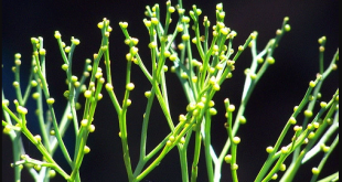
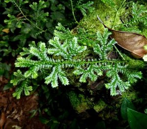
Selaginella sp. is the sole genus of the vascular plant under the family Selaginellaceae and order Selaginellales. The member of this order has herbaceous stems that are usually without any indication of secondary thickening. Commonly known as spike mosses.
Classification
- Division: Lycophyta
- Class: Ligulopsida
- Order: Selaginellales
- Family: Selaginellaceae
- Genus: Selaginella
Occurrence
Selaginella mostly occurs in tropical regions. It is worldwide in distribution. Many of them grow in damp forests. Approximately 40 species of Selaginella, found in temperate zones, are native to the United States. In tropical countries with moist and shady areas, around 700 species are found. Many of them grow in damp forests.
Some are xerophytic and grow on rocky cliffs or on soil that periodically becomes dry. This xerophytic species can withstand desiccation for months. A rosette-like system of branches coils tightly together as the plants dry out and re-expanding when it is moistened.
- Xerophytic: Selaginella lepidophylla (also called resurrection plant), Selaginella pilifera
- Epiphytic: Selaginella oregano
Some other commercially available species are-
- Red Selaginella – Selaginella erythropus
- Golden Selaginella – Selaginella kraussiana
- Peacock fern – Selaginella uncinata
- Absorbtive fern – Selaginella braunii
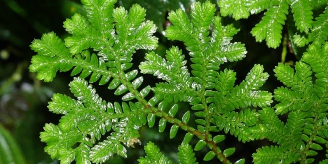
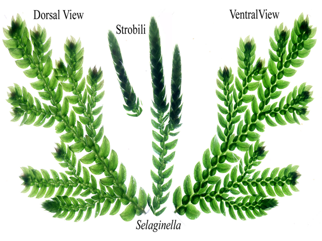
External Features of Selaginella
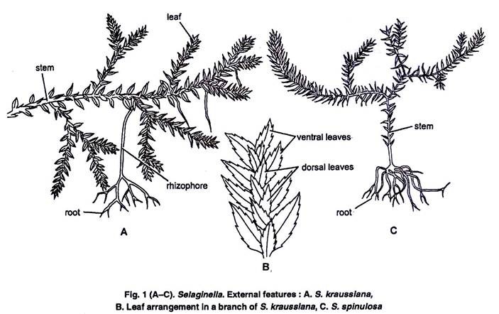
Stem
The stem is typically dichotomous often monopodial, others are climbing or suberect. The majority are perennial and few are delicate annuals. They vary in length from a few centimeters to several meters.
S. kraussiana shows a single layer of epidermal prosenchymatous fibers without any stomatal opening. Below it is the cortex of angular parenchymatous cells usually without intercellular spaces. The outer cortex cells are thickened in some, especially, xerophytic species.
The vascular cylinder in Selaginella species varies from protostelic to siphonostelic and monostelic to polystelic (with 2 to 16 separate steles). But, on the whole, the stelar arrangement is simpler than in Lycopodium. S. kraussiana is distelic.
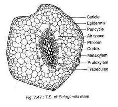
In the stele there is an exarch xylem at the center, the metaxylem shows scalariform tracheids, and a protoxylem lies towards the circumference. The xylem is surrounded by one or two layers of parenchyma which again is surrounded by a single layer of sieve tubes with sieves on lateral walls forming the phloem.
Leaves
Based on the position of the leaves, the genus Selaginella is divided into two subgenera-
- Homoeophyllum
- Heterophyllum
Homoeophyllum: The plant body is upright and has leaves arranged in a spiral pattern that are more or less uniform.
Heterophyllum: the dorsiventral and prostrate plant body is present with distinct heterophylly of leaves including the majority of species. In this subgenus, the leaves are two types. Two rows of large ventral leaves and two rows of small dorsal leaves.
A t.s. of the leaf shows a simple organization. At the center, there is a simple vascular bundle. The epidermis is one layer thick and the stomata are usually present on the lower epidermis only. The mesophyll is uniformly formed either of all spongy or all palisade-like elongated cells full of air spaces. Mesophyll cells have single cup-shaped or many chloroplastids according to species.
In all cases, there are several spindle-shaped, pyrenoid-like bodies at the center of each chloroplast. The bodies may be trans-through ligules at the base of the leaf, formed into rudimentary starch grains. The only other Embryophyte with similar chloroplasts is Anthocerotopsida.
Legule
On the upper surface of each leaf near the base small flap-like appendages are present called ‘Legule’.t
In sporophylls, legule lies between the sporangium and the leaf blade. it becomes prominent and functional during the growth of leaves. In its maturity the legule withers away.
The definite function of the legule is unknown but it’s predicted that it may act as water absorbing or water-secreting body.
Rhizophore
At the place of branching the stem or at the prostrate axis arises an elongated downwardly growing, colorless, leafless cylindrical appendages called rhizophore. Each of these typically develops a tuft of adventitious roots at the free end.
This structure provides support to weak stems.
Root
All roots borne by adult plants are adventitious in origin. In most species, rhizoids are formed only at the distal end of the rhinophores. However, in a few species like S. wallichii, adventitious roots can develop anywhere along the stem.
The roots of Selaginella species are delicate, sparsely branched, and have a small stele.Typically this is a monarch in an organization and has one phloem and one xylem mass. Primary roots are of a short-lif
Reproduction
Vegetative reproduction takes place by
- Fragmentation
- Stems or tubers
- Bulbills
Asexual reproduction
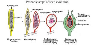
asexual reproduction takes place by producing spores. here spores are heterosporous which means produce two types of spores.
In S. patula, Microsporophyll and Megasporophyll are grouped together in strobiles and cones at the top of stems or lateral branches. The plant also produces shoots with sporophyll and foliage leaves in an alternating pattern along the stems.
Most commonly both sporophylls are borne in the same strobilus
E.g. S. kraussiana, S. helvetica
Sporophylls may be born in different strobila.
E.g. S. gracilis
In S. rapestris and S. selagenoides the upper portion may act as megasporangium and the lower portion may act as microsporangium.
In S.kraussiana one megaspore remains at the base and the others are microphyllous.
Microsporangium is on one side and megasporangium on another side in S. inaquaefoiia.
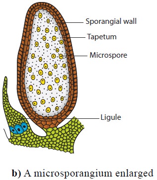
Development of sporangium in Selaginella
early development of sporangium similar to the spore mother cell. here the development of sporangium is of eusporangiate type. The sporangium arises from initial cells of the axis, however, at maturity, the sporangium comes to lie in the axile of the sporophyll and then it looks as if it had originated right on the sporophyll. , unlike Lycopodium sp tapetum derives from sporogenous tissues rather than the jacket layer.
Right from stage 4 onwards sporangia start differentiating into two types of spores – microspores and megaspores.
Microspore mother cells through meiosis form microspore tetrad. in a megaspore, all the megaspore mother cells degenerate before meiosis except one. this one divides meiotically to form a tetrad of haploid megaspores. Sometimes 8,12 or 16 megaspores may occur.
In S. rapestris, only one megaspore remains.
Development of Male gametophyte
Male gametophyte develops within the wall of the microspore before it is shed from the microsporangium. the ciliate antherozoids similar to bryophyte and lycopodium. microgametophyte is greatly reduced and is developed entirely within the wall of microspores.
Therefore the vegetative prothalli are not essential. When the antherozoids mature, the wall of the microspore splits open along the triradiated ridge, freeing the antherozoids.
Development of megagametophyte
The germination of megagametophytes within megasporangium differs in different species.
The megagametophyte in S. kraussiana is released when the first archegonium is formed, whereas in S. rapestris, the megaspore remains within the megasporangium until fertilization or even after the development of the embryo.
The megaspore nucleus divides repeatedly forming three nuclei. the nuclei in young gametophytes lie in the peripheral layer of cytoplasm surrounding a large central vacuole.
The nuclei increase, causing the cytoplasm to thicken and the vacuole to fill with cytoplasm. The formation of the wall around the daughter nuclei starts first at the apical part of the spore, creating a cushion of cellular tissue.
In some species, the apical cushion lies in continuation with the rest of the prothallus. but in some species separated from the rest by the diaphragm below within which 3 nuclei occur. Few archegonial initials make their appearance in the apical tissues. shortly after the spore wall breaks the apical cushion protrudes. the basal non-cellular region becomes filled with large cells. these cells act as a food reserve. Exposed apical regions give off few rhizoids.
Fertilization may take place while the megagametophyte is still with the megasporangium or after falling or remaining attached to the megasporangium for some time the falls to the ground, where primary rhizospheres develop roots into the soil and grow into the soil and start independent lifecycle.
The New Sporophyte
The first division of the zygote is usually transverse. The upper cell gives rise to one or more celled suspensors and the lower cell to the embryo. The embryo initial divides vertically The two resultant cells again divide vertically to form four cells one of which cuts off the stem initial from its base
Gradually, the embryo organizes into sectors on one side of which is the stem between two cotyledons, on the other side is the foot (for sucking food material from the endosperm) occupying a major part of the base, and the rhizophore initial on the top of it
The life cycle of Selaginella sp

References
Class Note