Periderm is the corky outer layer of a plant stem formed in secondary thickening or as a response to injury or infection. It is a cylindrical tissue that covers the surfaces of stems and roots of perennial plants during early secondary growth; therefore it is not found in monocots and is confined to those gymnosperms and eudicots that show secondary growth.
In this article, Periderm, it’s structure and development will be discussed briefly.
Periderm
Due to the continued formation of secondary tissues, in the older stem and roots, however, the epidermis gets stretched and ultimately tends to rupture and followed by the death of
epidermal cells and outer tissues and a new protective layer is developed called a periderm.
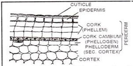
Structure of periderm
Periderm consists of three parts:
- The meristem is known as phellogen or cork cambium.
- The layer of cells cut off by phellogen on the outer side constitutes a phellem or cork cell.
- The layer of cells cut off by the phellogen towards the inner side constitutes phelloderm.
These three parts are described as follows:
1. Phellogen
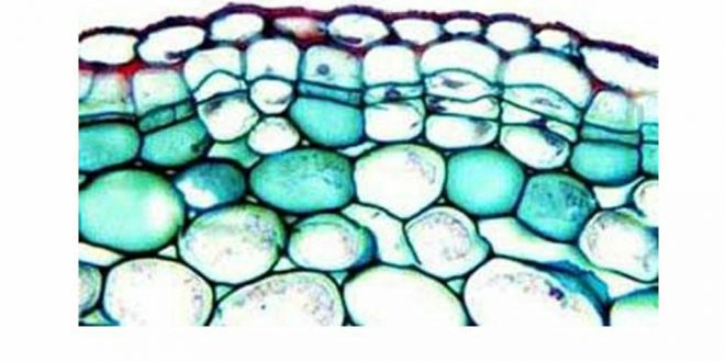
Phellogen is a type of lateral meristem. In contrast to the vascular cambium, the phellogen is relatively simple in structure.
- Phellogen arises in the epidermis, hypodermis, cortex, and phloem tissue.
- Phellogen is composed of one type of cell. Cells are rectangular in cross-section and radially flattened.
- Protoplasm contains tannins and chloroplast.
- Intercellular spaces are lacking except in lenticels.
- Due to the activity of the phellogen plant axis increases in thickness.
- Phellogen divides by periclinal division and produced phellem or cork cells on
the outside and phelloderm on the inner side.
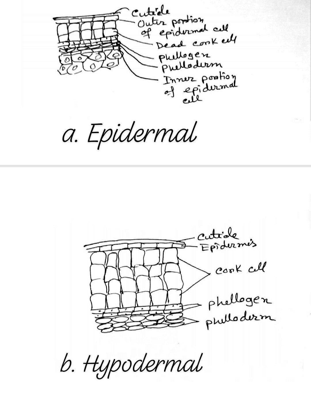
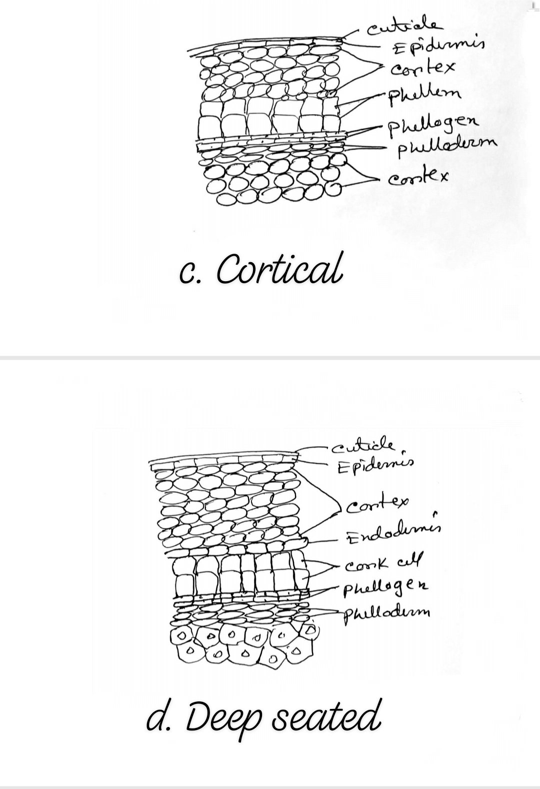
Place of origin of phellogen
- In most stems, the first phellogen arises in the subepidermal layer. In a few plants, the phellogen arises in the epidermal cells (Nerium, Pyrus). Sometimes only a part of the phellogen is developed from the epidermis while the other part arises in subepidermal cells (Pyrus). In some stem, the second and third cortical layer initiates the development of periderm (Robinia, Aristolochia). In still other plants the phellogen arises near the vascular region or directly in the phloem (Punica, Vitis).
- At the time of the beginning of the development of a phellogen in epidermal cells, the protoplast loses central vacuoles and the cytoplasm increases in amount and becomes more richly granular. As soon as this initial layer develops, it divides tangentially and to a lesser extent radially, in a similar way as division takes place in the cambium.
- Generally several to many times as many cells are cut off towards the outside (phellem) as towards the inside (phelloderm). Phelloderm cells are few or absent. Rarely phelloderm is greater in amount than phellem.
The origin and development of Phellogen Diagrams are given below:
2. Phellem (Cork cells)
The cells that constitute phellem are commonly known as cork cells. They are like the phellogen from which they are derived.
- Cells are dead at a mature stage.
- There are no intercellular spaces in cork cells.
- Suberin is present in the cell wall. In some cells, suberin is absent called phelloid cells.
- The wall of the cork cell may be colorless or a colored substance may be present in the cell lumen.
- As seen in the tangential section, cork cells are polygonal and uniform in shape, often radially thin as seen in the cross-section of the stem.
- The cells of commercial cork (Quercus suber) are radially elongated as seen in the transverse section.
- In the periderm of the Betula and Prunus, the cork cells are elongated tangentially as seen in the cross-section.
3. Phelloderm
The phellogen cuts off the phelloderm cells towards the inner side which are living cells with
cellulose walls.
- The cells are loosely arranged and pitted.
- In most plants, they resemble cortical cells in the wall structure and contents.
- Their shape is similar to that of phellogen cells.
- They may be distinguished from cortical cells by their arrangement in radial series resulting from their origin from the tangentially dividing phellogen.
- In some species, they act as photosynthetic tissue and aid in starch storage.
- They are pitted like other parenchyma cells.
- Occasionally the sclereids and other such specialized cells occur in phelloderm.
- The term secondary cortex is sometimes applied to phelloderm which does not see to be appropriate.
Commercial cork
The development of the periderm layers in the cork Oaks (Quercus suber) is of special interest. The ability of the plant to produce phellogen in deeper layers, when the superficial periderm is removed, is utilized in the production of commercial cork (Q. Suber). At the age of about twenty years, when the tree is about 40 cm in circumference, this outer layer, known as virgin cork is removed by stripping to the phellogen.
Reference:
1. Class Lecture of Parveen Rashid, PhD
Professor, Department of Botany, University of Dhaka.
 Plantlet The Blogging Platform of Department of Botany, University of Dhaka

This article is ranked on the second position of google for the keywords ‘periderm structure’, ‘periderm structure and function’, ‘periderm plant diagram’. Congrats!
Try to add more relevant informations.